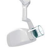
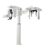
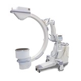
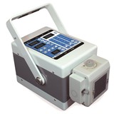
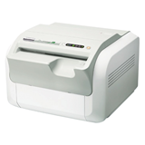
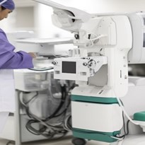
Mobile X-ray machines play a pivotal role in modern healthcare, offering the convenience of on-the-go diagnostic imaging. However, ensuring the safety and quality of these mobile imaging systems is of paramount importance. In this article, we delve into the key aspects that healthcare professionals and institutions must consider to guarantee the highest standards of safety and quality in mobile X-ray imaging.
I. Compliance with Radiation Safety Regulations
A. Regulatory Framework
- Regulatory Bodies: Mobile X-ray machines, like all medical imaging equipment, are subject to stringent regulations and oversight by various regulatory bodies. These agencies, such as the Food and Drug Administration (FDA) in the United States, the European Medicines Agency (EMA) in Europe, and their counterparts worldwide, are responsible for ensuring that these devices meet safety and performance standards.
- Relevant Regulations and Guidelines: Within this regulatory framework, there exist a plethora of regulations and guidelines that govern the design, manufacture, and use of mobile X-ray machines. These regulations often specify radiation safety standards, quality control measures, and operator training requirements. It is imperative that healthcare providers are well-versed in these regulations to ensure compliance.
B. Equipment Compliance
- Importance of Compliance: Compliance with radiation safety regulations is not a mere formality; it is a fundamental requirement to ensure the safety of both patients and healthcare providers. Non-compliant equipment can pose significant risks, leading to inaccurate diagnoses and potential harm due to excessive radiation exposure.
- Factors to Consider: When selecting mobile X-ray equipment, several factors must be considered to ensure compliance. These factors encompass the machine's design, radiation shielding, dose control mechanisms, and the presence of essential safety features. Choosing equipment that aligns with regulatory standards is the first step toward a safe and effective mobile X-ray imaging program.
II. Proper Shielding and Radiation Protection Measures
A. Shielding Requirements
- Shielding in mobile X-ray machines is imperative to ensure the safety of both operators and patients.
- Various shielding materials are employed in mobile X-ray machines, each selected for its unique properties and effectiveness.
- Lead: Lead is a commonly used shielding material due to its high density, which effectively absorbs and attenuates X-rays. It is often used to line the interior of X-ray tubes and surrounding structures to prevent radiation leakage.
- Bismuth: Bismuth is another material known for its radiation-absorbing properties. It is used in some portable shielding barriers and aprons worn by healthcare providers.
- Composite Materials: Modern shielding solutions may incorporate composite materials that combine lead or bismuth with other materials to achieve optimal radiation attenuation while reducing weight and bulk.
B. Radiation Protection Protocols
- Maintaining safe distances during mobile X-ray imaging procedures is a fundamental aspect of radiation protection.
- Operators and other personnel should adhere to recommended minimum distances from the X-ray source and the patient whenever possible.
- This minimizes their radiation exposure and reduces the risk of potential harm.
- Personal Protective Equipment (PPE) plays a crucial role in shielding operators from radiation.
- Lead aprons, thyroid collars, lead gloves, and protective eyewear are examples of PPE designed to minimize radiation exposure to specific areas of the body.
- Proper usage and regular inspection of PPE are essential to ensure their effectiveness.
III. Training and Certification for Mobile X-Ray Operators
A. Operator Training
- Proper training for mobile X-ray operators is of utmost importance to ensure the safe and accurate use of these medical imaging devices.
- Operator training programs are designed to equip healthcare professionals with the necessary knowledge and skills to handle mobile X-ray equipment effectively.
- Topics covered in operator training programs typically include:
- Radiation Physics and Safety: Operators learn the fundamental principles of radiation, its biological effects, and safety measures to minimize exposure.
- Equipment Operation: Comprehensive training on the operation of mobile X-ray machines, including setting exposure parameters, positioning patients, and image acquisition.
- Patient Care and Communication: Ensuring operators are skilled in patient interactions, positioning techniques, and addressing patient concerns related to radiation.
- Infection Control: Proper sterilization and hygiene practices to prevent the spread of infections during mobile X-ray procedures.
- Emergency Procedures: Training on how to respond to emergencies, such as equipment malfunctions or adverse reactions.
B. Certification and Competency
- Mobile X-ray operators must undergo certification and competency assessments to demonstrate their proficiency in using the equipment safely and effectively.
- Certification is a formal recognition of an operator's qualifications and competence in mobile X-ray imaging.
- The need for operator certification is driven by several key factors:
- Patient Safety: Certified operators are more likely to follow best practices and safety guidelines, reducing the risk of errors that could harm patients.
- Regulatory Compliance: Many regulatory bodies require operators to be certified as part of compliance with radiation safety regulations.
- Quality Assurance: Certification ensures that operators can consistently produce high-quality diagnostic images, which is crucial for accurate diagnoses and treatment planning.
- The certification process typically involves:
- Completion of an accredited training program.
- Passing a written examination that tests knowledge of radiation safety, equipment operation, and relevant regulations.
- Demonstrating practical skills in operating mobile X-ray machines, including patient positioning and image acquisition.
- Furthermore, ongoing competency assessments are essential to ensure that certified operators maintain their skills and stay updated with advancements in mobile X-ray technology and safety protocols. These assessments may include periodic re-certification, continuing education, and performance evaluations
IV. Quality Control and Image Diagnostic Accuracy
A. Quality Control Measures
Quality control plays a pivotal role in ensuring the reliability and safety of mobile X-ray imaging.
Routine quality assurance tests and checks are indispensable components of this process.
- Importance of Quality Control: Quality control, in the context of mobile X-ray imaging, is a systematic approach aimed at maintaining the consistency, accuracy, and safety of radiographic images. It encompasses a set of measures and protocols designed to detect and rectify issues that may compromise the quality of images or pose risks to patients and operators.
- Routine Quality Assurance Tests: To uphold the highest standards in mobile X-ray imaging, healthcare providers must implement a regimen of routine quality assurance tests and checks. These tests are conducted on both the equipment and the imaging process itself. They serve several essential purposes:
- Equipment Performance Assessment: Regular assessments of X-ray machines are conducted to ensure they are functioning within specified parameters. This includes evaluating factors such as exposure accuracy, tube output, and collimation precision.
- Image Consistency: Quality control procedures verify the consistency of images produced over time. This is crucial in diagnosing medical conditions accurately, as variations in image quality can lead to misinterpretations.
- Patient Safety: Quality control measures also include evaluating radiation exposure levels to confirm that they are within safe limits for patients. This involves monitoring radiation dose indicators and assessing any potential risks.
- Operator Competence: Quality control extends to operator competence and adherence to safety protocols. Regular audits and assessments of operators' practices ensure they are using the equipment correctly and following safety guidelines.
- Equipment Performance Assessment: Regular assessments of X-ray machines are conducted to ensure they are functioning within specified parameters. This includes evaluating factors such as exposure accuracy, tube output, and collimation precision.
B. Image Diagnostic Accuracy
Accurate diagnostic images are the cornerstone of effective patient care in mobile X-ray imaging.
Multiple factors influence image quality and accuracy, each requiring careful consideration.
- The Significance of Accuracy: Accurate diagnostic images are essential for guiding clinical decisions and treatment plans. Even minor discrepancies in image quality can lead to incorrect diagnoses or incomplete assessments of a patient's condition, potentially compromising their health outcomes.
- Factors Affecting Image Quality and Accuracy
- Exposure Parameters: Proper selection of exposure factors such as tube current, voltage, and exposure time is crucial. Incorrect settings can result in overexposed or underexposed images with reduced diagnostic value.
- Patient Positioning: The positioning of the patient and the X-ray equipment must be precise to capture the targeted anatomical area accurately. Misalignment can lead to distortion or inadequate visualization.
- Motion Artifacts: Patient motion during the imaging process can introduce blurriness or artifacts in the images. Techniques such as immobilization and patient cooperation are essential to minimize these issues.
- Image Processing: Post-processing techniques, including image enhancement and manipulation, should be used judiciously. Over-processing can introduce artifacts and alter the original image's integrity.
- Image Display and Interpretation: The quality of monitors and viewing conditions can impact the interpretation of diagnostic images. Ensuring optimal monitor calibration and ambient lighting is vital.
- Radiation Dose Optimization: Balancing the need for image quality with minimizing radiation dose to patients is a critical consideration. Utilizing appropriate dose reduction techniques and ALARA (As Low As Reasonably Achievable) principles is essential.
VI. Minimizing Radiation Exposure for Patients and Healthcare Providers
Radiation exposure is a significant concern in medical imaging, especially when it comes to mobile X-ray procedures. Ensuring patient safety is paramount, and healthcare providers must adopt strategies to minimize radiation exposure during these examinations. In this section, we will discuss various approaches and considerations for safeguarding patients from unnecessary radiation risks.
A. Patient Risk Assessment
Before delving into strategies for minimizing radiation exposure, it's crucial to emphasize the importance of patient risk assessment. Every patient is unique, and their medical condition may dictate the necessity and frequency of X-ray imaging. Healthcare providers must conduct a thorough assessment to determine whether an X-ray procedure is justified for a particular patient. Here are key points to consider:
- Clinical Indication: The foremost consideration is whether the X-ray is clinically justified. Is there a clear medical need for the imaging, such as diagnosing a specific condition or monitoring treatment progress?
- Alternative Imaging Modalities: In some cases, alternative imaging modalities like ultrasound or MRI may provide the necessary diagnostic information without using ionizing radiation. Providers should explore these options when appropriate.
- Pregnancy: Special attention must be paid to pregnant patients. Ionizing radiation can pose risks to the developing fetus. If possible, X-ray procedures should be postponed until after pregnancy, except in emergencies where the benefits clearly outweigh the risks.
- Pediatric Patients: Children are more radiation-sensitive than adults, so minimizing exposure is especially critical for pediatric patients. Proper dose adjustment and pediatric-specific protocols should be used.
- Cumulative Exposure: Consider a patient's cumulative radiation exposure from previous imaging procedures. Avoid unnecessary repeat exams and ensure that the cumulative dose remains within safe limits.
B. Strategies for Minimizing Radiation Exposure to Patients
- Utilization of Proper Equipment
The choice of X-ray equipment is foundational in minimizing patient radiation exposure. Modern mobile X-ray machines offer advanced features like dose optimization and image enhancement. Opt for equipment that incorporates these technologies to reduce the radiation dose required for accurate imaging.
- Optimization of Exposure Parameters
Radiologic technologists should be trained to adjust exposure parameters such as tube current, voltage, and exposure time according to the specific imaging needs and the patient's size. This optimization ensures that only the necessary radiation is used.
- Patient Positioning and Immobilization
Proper patient positioning and immobilization techniques are crucial. They help ensure that the X-ray beam is precisely targeted at the area of interest, reducing the need for retakes and additional exposures.
- Filtration and Collimation
Employing appropriate filters and collimators narrows the X-ray beam to the desired area and filters out unnecessary radiation. This focused approach significantly reduces patient exposure.
- Pulse Mode Imaging
Pulse mode imaging, available in many modern mobile X-ray systems, allows for the emission of X-rays in short, controlled bursts. This limits the total radiation exposure while maintaining image quality.
- Lead Aprons and Thyroid Shields
Utilize lead aprons and thyroid shields for patients, especially in cases where the area to be imaged is far from vital organs. These protective shields provide an additional layer of safety by shielding sensitive tissues.
- Pediatric Imaging Protocols
Pediatric patients are more sensitive to radiation. Implementing specific pediatric imaging protocols, including lower dose settings and specialized techniques, helps minimize their exposure while obtaining diagnostically useful images.
- Pregnancy Assessment
Special consideration should be given to pregnant patients. Conduct a thorough risk assessment and ensure that the benefits of the X-ray examination outweigh the potential risks to the fetus. Whenever possible, alternative imaging modalities like ultrasound or MRI should be considered.
B.The Importance of Patient Risk Assessment
Patient risk assessment is a fundamental aspect of radiation safety in mobile X-ray imaging. It involves evaluating the potential risks and benefits associated with a specific X-ray examination for an individual patient. By tailoring the imaging approach to the patient's unique circumstances, healthcare providers can minimize radiation exposure while maintaining diagnostic accuracy.
Key Components of Patient Risk Assessment
- Clinical Indication
Before proceeding with an X-ray examination, healthcare providers must assess the clinical indication. Is the examination necessary for diagnosis or treatment planning? Is there an alternative imaging modality that does not involve ionizing radiation?
- Patient Characteristics
Factors such as age, gender, medical history, and pregnancy status play a significant role in patient risk assessment. For instance, pediatric and pregnant patients are more susceptible to radiation-related risks and require specialized considerations.
- Imaging Protocol Selection
Based on the clinical indication and patient characteristics, healthcare providers should select the most appropriate imaging protocol. This includes determining the type of X-ray examination, exposure parameters, and any necessary shielding.
- Informed Consent
Informed consent is an essential aspect of patient risk assessment. Patients should be fully informed about the procedure, its risks, benefits, and alternatives. Their consent demonstrates their understanding and willingness to proceed.
- Documentation
Comprehensive documentation of the risk assessment process is crucial for accountability and quality assurance. This includes recording the clinical indication, patient characteristics, selected imaging protocol, and informed consent details.
In conclusion, ensuring safety and quality in mobile X-ray imaging is a paramount responsibility in modern healthcare. Adherence to radiation safety regulations, compliance with equipment standards, and the use of proper shielding and radiation protection measures are foundational steps to safeguard both patients and healthcare providers. Rigorous operator training and certification programs ensure that mobile X-ray operators are competent and capable of maintaining safety standards during procedures. Quality control measures, including routine testing and checks, guarantee the reliability and consistency of diagnostic images, contributing to accurate diagnoses. Additionally, minimizing radiation exposure for patients and healthcare providers through careful patient risk assessment and the implementation of dose-reduction strategies is essential for maintaining patient safety. By diligently addressing these key aspects, healthcare institutions can uphold the highest standards of safety and quality in mobile X-ray imaging, ultimately enhancing patient care and outcomes while minimizing potential risks.


-160x160-state_article-rel-cat.png)




-160x160-state_article-rel-cat.png)
-160x160-state_article-rel-cat.png)

