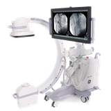

In the area of modern medical practice, technological advancements have played a pivotal role in enhancing patient care and surgical procedures. Among these advancements, X-ray machines stand as stalwarts, facilitating accurate diagnostics and guiding intricate surgical interventions. To harness the full potential of these machines, it is imperative that medical professionals undergo comprehensive training to navigate the learning curve associated with their operation. This pillar post delves into the intricacies of incorporating surgical loupes and headlights into the usage of X-ray machines, focusing on the training and adaptation required for seamless integration.
I. Learning Curve and Skill Development for New Users
Mastering the operation of an X-Ray machine requires a systematic approach that encompasses not only technical knowledge but also practical skills. As medical professionals venture into the realm of radiology and diagnostics, understanding the nuances of X-Ray machine operation becomes paramount. This section delves into the essential steps that constitute the learning curve for new users, encompassing everything from grasping the basics to navigating complex procedures with finesse.
A. Understanding the Basics of X-Ray Machine Operation
Before embarking on the journey of utilizing an X-Ray machine effectively, users must establish a solid foundation by comprehending its fundamental principles. At its core, an X-Ray machine functions as a sophisticated imaging tool that generates images by passing X-Ray radiation through the body, capturing variations in tissue density. It is imperative for users to grasp the mechanics of X-Ray generation, the interplay of radiation with the human body, and the intricate process of image formation.
B. Familiarization with Control Panel and Settings
A pivotal aspect of mastering X-Ray machine operation lies in becoming intimately acquainted with its control panel and settings. The control panel serves as the nerve center, allowing users to manipulate various parameters that influence image quality, radiation dosage, and exposure settings. Proficiency in adjusting these settings enables radiographers to tailor imaging procedures according to the unique anatomical requirements of each patient, ensuring accurate diagnosis while minimizing radiation exposure.
C. Practice in Simulated Environments for Skill Refinement
Skill refinement is a cornerstone of mastering any complex apparatus, and the X-Ray machine is no exception. Novice users are strongly encouraged to engage in simulated environments that replicate real-world scenarios. These environments provide a controlled platform for practicing positioning techniques, fine-tuning exposure settings, and perfecting the alignment of the X-Ray beam with the targeted anatomy. Such deliberate practice not only hones technical skills but also fosters the development of a confident and composed approach to handling the machine during actual clinical settings.
D. Importance of Proper Hand-Eye Coordination
Effective operation of an X-Ray machine relies heavily on the synergy between hand-eye coordination and cognitive understanding. Radiographers must seamlessly translate their understanding of anatomical structures into precise positioning of patients and X-Ray equipment. This demands a delicate balance between manual dexterity, visual acuity, and the ability to navigate the intricate choreography of machine manipulation. As users cultivate their hand-eye coordination, the quality and accuracy of generated images are significantly enhanced.
E. Gradual Progression from Simple to Complex Procedures
Mastery over X-Ray machine operation is an incremental process that evolves through a series of steps, gradually advancing from simpler procedures to more intricate ones. Beginners should commence their journey by undertaking routine imaging tasks that involve relatively straightforward anatomical regions. As users gain confidence and proficiency, they can progressively tackle more complex procedures, such as those involving weight-bearing joints or specialized radiographic techniques. This incremental approach ensures that users build a robust skill set while continually expanding their repertoire of techniques.
II. Overcoming Depth Perception and Field of View Adjustments
X-ray imaging, with its unparalleled ability to reveal internal structures, has revolutionized medical diagnostics and interventions. However, the nature of X-ray imaging presents specific challenges related to depth perception and the limited field of view. Surgeons and medical professionals working with X-ray machines must navigate these obstacles to ensure accurate and effective procedures. This section delves into the intricacies of overcoming depth perception challenges and making the most of the often-restricted field of view, utilizing tools such as surgical loupes and headlights.
A. Challenges Associated with Depth Perception in X-Ray Imaging
- Layered Structures: X-ray images provide a two-dimensional representation of three-dimensional structures, leading to potential confusion in differentiating overlapping structures.
- Loss of Depth Cues: Traditional depth cues, such as binocular disparity and motion parallax, are absent in static X-ray images, making accurate depth interpretation challenging.
- Complex Procedures: Surgeons conducting intricate procedures require precise depth perception to gauge distances and make informed decisions.
B. Adapting to Limited Field of View in X-Ray Scans
- Scope of Visualization: X-ray scans offer a limited view of the target area, requiring medical professionals to mentally reconstruct a comprehensive image from fragmented data.
- Spatial Orientation: Adjusting to the orientation of structures within the limited field of view demands a profound understanding of anatomy and spatial relationships.
- Risk of Misinterpretation: Incomplete information due to limited visualization can lead to misdiagnoses or missed critical details.
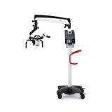
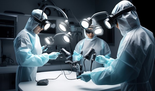
C. Utilizing Surgical Loupes to Enhance Depth Perception
- Magnification Advantage: Surgical loupes provide magnification, aiding in distinguishing subtle differences in depth and structures that might be indistinguishable with the naked eye.
- Improved Visual Acuity: Enhanced magnification sharpens details, allowing surgeons to identify fine anatomical features that contribute to accurate depth perception.
- Skill Development: Proficient use of loupes demands practice and training to synchronize magnified visuals with manual dexterity.
D. Integrating Headlights for Improved Visual Clarity
- Targeted Illumination: Headlights direct light precisely where needed, minimizing shadows and enhancing visibility of the surgical field during X-ray-guided procedures.
- Enhanced Contrast: Proper lighting reduces image obscurity caused by overexposure or underexposure, aiding in clear interpretation of X-ray images.
- Adjustable Intensity: Surgeons can control the intensity of headlights to balance illumination with X-ray imaging requirements.
E. Strategies to Overcome Distortions and Visual Limitations
- Reference Landmarks: Relating X-ray images to anatomical landmarks helps mitigate distortions and supports accurate mental reconstruction of the target area.
- Comparative Analysis: Comparing X-ray findings with other diagnostic imaging modalities, such as MRI or CT scans, offers a comprehensive understanding of the patient's condition.
- Interdisciplinary Collaboration: Involving radiologists and other specialists can provide valuable insights, cross-validating interpretations for confident decision-making.
III. Training Programs and Workshops for Efficient Usage
In the rapidly evolving landscape of medical technology, the seamless and efficient integration of advanced equipment plays a pivotal role in ensuring optimal patient care. The X-Ray machine, a cornerstone of modern medical diagnostics, demands not only technical proficiency but also a comprehensive understanding of its operation and utilization. To bridge this knowledge gap and empower healthcare professionals, manufacturers offer structured training programs and immersive workshops that cater to both novice and seasoned users. These educational initiatives go beyond the conventional user manual, providing invaluable hands-on experiences and insights into maximizing the potential of X-ray machines.
A. Structured Training Programs Offered by Manufacturers
Manufacturers of X-ray machines recognize the significance of comprehensive training in harnessing the full capabilities of their equipment. In response to this need, they have designed structured training programs that cater to various skill levels. These programs encompass a wide array of topics, ranging from the fundamental principles of X-ray technology to the nuances of machine operation and troubleshooting.
These training programs typically include:
- Theoretical Modules: A solid theoretical foundation is essential for any successful endeavor, and operating an X-Ray machine is no exception. Training participants delve into the physics of X-Ray generation, radiation safety protocols, and the principles underlying image formation. This knowledge serves as a bedrock upon which practical skills are developed.
- Hands-on Practical Sessions: Theoretical knowledge gains real-world relevance through hands-on training sessions. Participants have the opportunity to operate the X-Ray machine under the guidance of experienced trainers. These practical exercises encompass a spectrum of scenarios, mimicking clinical situations, and offering a controlled environment for skill development.
- Safety Protocols and Regulatory Compliance: Given the importance of radiation safety, training programs emphasize adherence to safety protocols and regulatory guidelines. Participants gain an understanding of dose management, shielding techniques, and best practices to ensure patient and operator safety.
- Customization and Specialization: Manufacturers often tailor their training programs to cater to specific medical specialties. Whether it's radiology, orthopedics, dentistry, or any other field, the training content is customized to address the unique requirements of each domain.
B. Hands-on Workshops for Practical Experience
The transition from theoretical knowledge to practical application is a critical phase in the learning journey of healthcare professionals. Hands-on workshops provide an immersive environment for participants to consolidate their understanding and refine their skills. These workshops extend beyond the traditional classroom setup and simulate real clinical scenarios, creating an authentic learning experience.
Key features of hands-on workshops include:
- Scenario-Based Learning: Workshops are structured around scenarios that healthcare professionals commonly encounter in their practice. These scenarios incorporate the use of X-Ray machines to diagnose and address medical conditions, enhancing the participants' ability to make informed decisions.
- Collaborative Learning: Workshops often encourage collaboration among participants from diverse medical backgrounds. This fosters knowledge exchange, allowing participants to learn not only from trainers but also from their peers' insights and experiences.
- Realistic Simulations: High-fidelity simulations replicate clinical settings, complete with patient mannequins and anatomically accurate phantoms. This realism enables participants to familiarize themselves with patient positioning, machine adjustments, and image capture techniques.
- Immediate Feedback: Workshops provide a unique opportunity for immediate feedback from experienced trainers. This feedback loop accelerates the learning process, enabling participants to correct mistakes and refine their techniques in real time.
C. Role of Experienced Mentors in Skill Transfer
The guidance of experienced mentors significantly contributes to the effectiveness of training programs and workshops. These mentors, often seasoned radiographers or medical professionals with extensive experience in X-Ray technology, bring a wealth of practical insights to the learning process.
The role of mentors encompasses:
- Demonstration of Best Practices: Mentors showcase optimal techniques for machine setup, patient positioning, and image acquisition. Their demonstrations serve as benchmarks for participants to emulate in their own practice.
- Guidance in Problem-Solving: X-Ray imaging can present challenges, such as suboptimal image quality or technical glitches. Mentors leverage their experience to guide participants in troubleshooting and resolving these issues, ensuring uninterrupted clinical workflow.
- Contextual Application: Seasoned mentors contextualize theoretical concepts by sharing real-world anecdotes and case studies. This bridges the gap between theory and practice, helping participants grasp the practical implications of their learning.
- Addressing Advanced Scenarios: Advanced users seeking to refine their skills benefit from mentors who delve into intricate aspects of X-Ray imaging. These advanced workshops explore topics like image enhancement algorithms, contrast optimization, and specialized imaging techniques.
D. Incorporating Surgical Loupes into Training Modules
The integration of surgical loupes into X-Ray machine training modules enhances the educational experience for healthcare professionals. Surgical loupes, with their magnification capabilities, empower practitioners to visualize intricate details with enhanced clarity. Integrating their usage into training programs allows participants to understand the symbiotic relationship between X-Ray imaging and enhanced visualization.
Key considerations when incorporating surgical loupes into training include:
- Loupes Selection: Training modules guide participants in selecting appropriate loupes with suitable magnification levels for their specific medical tasks. This decision-making process ensures that practitioners strike the right balance between magnification and field of view.
- Ergonomic Integration: Proper ergonomic integration of surgical loupes is crucial to prevent discomfort during prolonged usage. Training programs emphasize the correct adjustment and alignment of loupes to maintain a natural posture while operating the X-Ray machine.
- Case-Based Learning: Practical scenarios where surgical loupes significantly contribute to diagnostic accuracy or procedural precision are highlighted. This practical context aids participants in recognizing opportunities where loupes can be seamlessly integrated into their workflow.
E. Addressing Common User Concerns and Queries
Training programs and workshops serve as platforms to address the common concerns and queries that healthcare professionals may have regarding X-Ray machine usage. These concerns may range from radiation exposure to image artifacts and everything in between. By providing expert insights and evidence-based answers, training initiatives equip participants with the knowledge to navigate potential challenges.
Common concerns addressed include:
- Radiation Safety: Participants are educated about radiation dose management and the measures in place to minimize patient and operator exposure. Clear explanations about how modern X-Ray machines optimize image quality while keeping radiation levels within safe limits are provided.
- Image Artifacts: Understanding and troubleshooting image artifacts are pivotal skills for any X-Ray operator. Training sessions elucidate the root causes of common artifacts and equip participants with strategies to mitigate or eliminate them.
- Optimizing Image Quality: Participants learn techniques to optimize image quality, considering factors like patient positioning, exposure settings, and patient cooperation. Practical tips for achieving diagnostic-quality images are shared.
- Workflow Efficiency: Addressing workflow challenges, such as patient throughput and equipment utilization, forms an integral part of training programs. Participants gain insights into streamlining processes without compromising on image quality.
In conclusion, the integration of X-ray machines with surgical loupes and headlights has the potential to redefine medical diagnostics and interventions. However, realizing this potential hinges on the comprehensive training and adaptation of healthcare professionals. Through structured training programs, hands-on workshops, experienced mentors, and the incorporation of surgical loupes, medical practitioners can harness the full capabilities of these advanced technologies, ultimately enhancing patient care and safety.