
Introduction
The study of cellular morphology is fundamental to understanding various physiological and pathological processes in the human body. In the early days of cytology, examining individual cells under a microscope presented numerous challenges, particularly in preserving cell integrity and obtaining representative samples. The advent of cytocentrifugation addressed these challenges, paving the way for more accurate and comprehensive cellular analysis.
Historical Evolution
The concept of centrifugation dates back to the late 19th century, but the specific application of centrifugal force to create monolayer cell preparations gained prominence in the mid-20th century. The first cytocentrifuge was developed by Albert H. Coons and Sydney M. Wentworth in the 1950s, marking a significant milestone in the field of cytology.
Early cytocentrifuges were rudimentary compared to contemporary models, but they laid the foundation for advancements that followed. Over the decades, technological innovations, such as the integration of computerized controls, improved rotor designs, and enhanced staining techniques, have refined the capabilities of cytocentrifuges.
Mechanism of Action
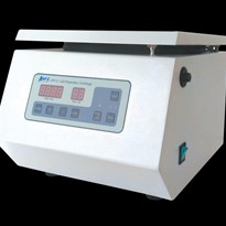
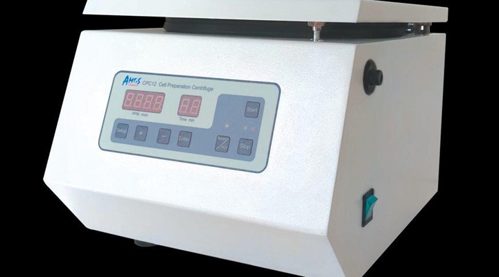
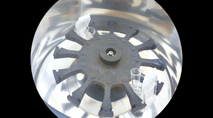
At its core, a cytocentrifuge employs centrifugal force to deposit cells onto a microscope slide in a monolayer fashion. The basic components include a rotor, sample chamber, and a slide holder. As the rotor spins, cells in suspension are forced against the slide, creating a uniform distribution of cells for subsequent analysis.
The ability to generate monolayer preparations is a key feature of cytocentrifugation. Unlike traditional methods that may result in clumped or overlapping cells, cytocentrifugation ensures a single layer of cells, allowing for clearer microscopic examination and more accurate cytological interpretation.
Types of Cytocentrifuges
Cytocentrifuges come in various designs, catering to different laboratory needs and applications. One common classification is based on whether the instrument is used for clinical or research purposes.
Clinical Cytocentrifuges:
These are designed for routine diagnostic procedures in clinical laboratories.
They are often automated and equipped with user-friendly interfaces to streamline the sample preparation process.
Clinical cytocentrifuges play a vital role in diagnosing conditions such as cancer, infections, and inflammatory diseases.
Research Cytocentrifuges:
Geared towards research laboratories and academic institutions, these instruments offer greater flexibility and customization.
Researchers often use them for studying cell biology, exploring novel staining techniques, and conducting experiments that require precise cell deposition.
Applications of Cytocentrifugation:
The versatility of cytocentrifuges has led to their widespread adoption across various scientific disciplines. Some key applications include:
Clinical Diagnostics
Cytocentrifugation is integral to clinical pathology, enabling the examination of cells for abnormalities associated with diseases such as cancer.
It plays a crucial role in the diagnosis of cytological disorders, including identifying malignant cells in body fluids.
Hematology
Blood cell differentials, a vital aspect of hematology, benefit from cytocentrifugation by providing a more accurate representation of cell populations.
Microbiology
In microbiology, cytocentrifugation aids in concentrating microorganisms from liquid samples, facilitating more effective identification and characterization.
Research in Cell Biology
Cytocentrifuges are extensively used in cell biology research, allowing scientists to study individual cells in a controlled environment.
The technology is invaluable for investigating cell structure, function, and interactions.
Stem Cell Research
Researchers working with stem cells leverage cytocentrifugation to prepare samples for analysis, contributing to advancements in regenerative medicine.
Immunology
Immunological studies benefit from cytocentrifugation, enabling researchers to analyze immune cells in various tissues and body fluids.
Challenges and Future Directions
While cytocentrifugation has significantly advanced cellular analysis, certain challenges persist. Achieving optimal cell recovery and maintaining cell viability during the centrifugation process are ongoing concerns. Researchers continue to explore innovations in rotor design, sample preparation techniques, and staining methodologies to address these challenges.
The future of cytocentrifugation holds promise with ongoing developments in automation, integration with other diagnostic technologies, and the potential for miniaturization. The advent of microfluidics and lab-on-a-chip technologies may further enhance the precision and efficiency of cytocentrifuges, opening new avenues for point-of-care diagnostics.
Conclusion
In conclusion, the cytocentrifuge stands as a cornerstone in the field of cellular analysis, offering unparalleled capabilities in preparing monolayer cell samples for microscopic examination. From its humble beginnings in the mid-20th century to the sophisticated instruments of today, cytocentrifuges have played a pivotal role in advancing diagnostics, research, and our understanding of cellular biology. As technology continues to evolve, the future holds exciting possibilities for further refining and expanding the applications of this indispensable laboratory tool.


-160x160-state_article-rel-cat.png)
-160x160-state_article-rel-cat.png)


-160x160-state_article-rel-cat.png)