
Unfortunately, if diabetic patients do not schedule and attend annual ophthalmologic appointments, the disease may go undetected, resulting in permanent vision loss or blindness. Historically, handheld fundus cameras have not been widely adopted for use in primary care-based diabetic retinopathy screening programs due to their relatively poor image quality and difficulty to operate. This study aims to test if a minimally-experienced operator can use the RetinaVue 100 Imager to successfully capture fundus images on a high percentage of patients, and if a high percentage of the images captured can be successfully interpreted by a remote retina specialist.
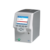
Study Objective: Is RetinaVue Network a viable solution for non-eye care settings (e.g primary care)? Can the RetinaVue 100 Imager be easily used to capture high-quality images on most people’s eyes
Participants were recruited using a convenience sample of employees who attended a health screening event. Participants had images of both eyes captured with the RetinaVue 100 Imager. All images were obtained in a nonmydriatic fashion (without the use of chemical pupil-dilating drops). Seven different camera operators participated in the study. Camera operators were trained*, but minimally experienced. Only two of the camera operators had a clinical background. Following image capture of both eyes, images from the RetinaVue 100 camera were instantly checked using the proprietary RetinaVue image quality algorithm, and then securely transferred to a board-certified, fellowship-trained retina specialist via the RetinaVue Network Software. The retina specialist from RetinaVue P.C. returned a diagnostic report for each subject, which included an assessment of any retinal, optic nerve, or systemic diseases identified and a recommended referral plan.
Study Outcomes
A total of 410 employees participated in the study. It took an average of four minutes to complete an exam. Overall, 92% of left eye images and 94% of right eye images could be read by a retina specialist. The camera immediately provides an indication of image quality—green scores denote good quality, which can be interpreted by a retina specialist, yellow scores denote acceptable quality which can most likely be read by a retina specialist, and red scores denote images that should be retaken. In this study, the image-quality score proved to be exceptionally accurate: 100% of the green (good quality) images were successfully interpreted by a retina specialist.
Additionally, abnormal pathology was found in 29 (7.21%) participants. Three participants required an immediate comprehensive eye examination by a specialist and another seven required urgent examinations for serious eye conditions. The remaining 19 patients required follow-up and monitoring by an ophthalmologist or retina specialist within the next six months. None of the ten participants requiring near-term assessment by an ophthalmologist or retina specialist were reported to be otherwise aware of problems with their eyes.
Conclusion
The RetinaVue 100 Imager can be effectively used in non-eye care settings (e.g. primary care offices) as a screening tool for diseases that present in the retina, including diabetic retinopathy. The camera proved easy to use as minimally experienced, non-clinical operators had a high success rate. An exam was completed in an average of just four minutes, proving that diabetic retinopathy screening using the RetinaVue 100 Imager can easily be added to a patient’s routine appointment. Image quality scoring provided by the camera provides accurate and instant feedback about whether the image needs to be retaken. In the sample of employees who participated in the study, a material number (7%) of potentially significant eye conditions requiring immediate follow-up were detected.
¹ CDC Vision Health Initiative (VHI), Common Eye Disorders. www.cdc.gov/visionhealth/basics/ced/index.html
² National Eye Institute, Facts about Diabetic Eye Disease.
https://nei.nih.gov/health/diabetic/retinopathy


-205x205.jpg)
-205x205.jpg)