When it comes to monitoring and understanding the heart's electrical activity, ECG machines play a crucial role in the medical field. These devices, also known as electrocardiographs, provide valuable insights into the heart's health and aid in diagnosing various cardiac conditions. In this article, we will explore the intricacies of ECG machines and electrocardiography, shedding light on their significance and functioning.
What is an ECG Machine?
Definition and Purpose
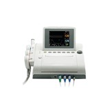
An ECG machine is a medical device used to record the electrical signals produced by the heart as it contracts and relaxes. These signals are translated into a visual representation known as an electrocardiogram. The primary purpose of an ECG machine is to detect irregularities in the heart's rhythm and identify potential cardiac issues.
How Does It Work?
An ECG machine works by using electrodes placed on the patient's skin to pick up the electrical signals generated by the heart. These electrodes are strategically positioned to record the electrical activity from different angles, providing a comprehensive view of the heart's performance.
Understanding Electrocardiography (ECG)
Definition and Explanation
Electrocardiography, commonly referred to as ECG or EKG (electrocardiogram), is the process of recording the heart's electrical activity. The ECG graph shows distinct waves, each representing a specific event during the cardiac cycle. By analyzing these waves, medical professionals can evaluate the heart's health and detect abnormalities.
The Electrical Activity of the Heart
The heart's electrical activity is divided into different phases: depolarization and repolarization. During depolarization, the heart's muscle contracts, and electrical impulses spread across the atria and ventricles. In contrast, repolarization is the period of relaxation when the heart is preparing for the next contraction.
Types of ECG Machines
-
3-Lead ECG
The 3-lead ECG is a basic type of ECG machine that uses three electrodes to monitor the heart's activity. It is commonly used in ambulatory settings and during routine check-ups to assess the heart's general condition.
-
12-Lead ECG
The 12-lead ECG is a more advanced version that utilizes 12 electrodes to provide a more detailed view of the heart's electrical signals. This type of ECG is extensively used in hospitals and clinics for comprehensive cardiac assessments.
-
Wireless ECG Monitors
Recent advancements in technology have led to the development of wireless ECG monitors, which offer convenience and mobility. These devices allow patients to perform ECG tests remotely and share the results with healthcare providers for analysis.
Importance of ECG Machines in Cardiac Diagnostic
Electrocardiography (ECG) machines are indispensable tools in the field of cardiac diagnostics, providing invaluable insights into the health and function of the heart. These medical devices play a crucial role in diagnosing various heart conditions and monitoring patients with known cardiac issues. Let's explore the significance of ECG machines in cardiac diagnostics and why they are an essential component of modern healthcare.
1. Detecting Irregularities in Heart Rhythm
One of the primary purposes of ECG machines is to detect irregularities in the heart's rhythm. An electrocardiogram records the electrical activity of the heart, represented by distinctive waves on the graph. These waves allow medical professionals to identify abnormal patterns, such as arrhythmias, which can be indicative of serious cardiac conditions.
2. Diagnosing Heart Attacks
ECG machines are critical in diagnosing heart attacks (myocardial infarctions). When a heart attack occurs, the blood flow to a part of the heart is blocked, resulting in damaged heart muscle. An ECG can detect characteristic changes in the heart's electrical activity, aiding in swift diagnosis and immediate intervention.
3. Evaluating Conduction Disorders
The heart relies on a well-coordinated electrical system to contract effectively and pump blood efficiently. Conduction disorders, such as atrial fibrillation or heart block, can disrupt this system. ECG machines can pinpoint such issues by analyzing the heart's electrical impulses and assist in devising appropriate treatment plans.
4. Monitoring Heart Health
Patients with known heart conditions or those at risk of heart problems require regular monitoring. ECG machines provide a non-invasive and efficient means of tracking the heart's performance over time. Physicians can use the data from multiple ECG tests to assess the effectiveness of treatments and make necessary adjustments.
5. Preoperative Evaluation
Before certain surgical procedures, especially those involving anesthesia, it is crucial to evaluate the patient's heart health. ECG machines play a vital role in preoperative assessments, helping medical teams ensure that the patient can undergo surgery safely.
6. Assessing Response to Medication
For patients on heart medications, ECGs are essential in assessing the body's response to the treatment. By monitoring changes in the heart's electrical activity, healthcare providers can gauge the effectiveness of the prescribed medication and make changes as needed.
7. Screening for Cardiac Disorders
In some cases, individuals may have undiagnosed cardiac conditions or be at risk due to family history or lifestyle factors. ECG machines can be used for screening purposes, enabling early detection and intervention, which is crucial for preventing further complications.
8. Quick and Non-invasive Testing
ECG tests are quick and non-invasive, making them highly convenient for both patients and medical professionals. The procedure involves attaching electrodes to the patient's skin, and the results are available almost instantly. This efficiency is vital in emergency situations when immediate decisions need to be made.
9. Aid in Treatment Decisions
The information gathered from ECG tests provides valuable data that helps physicians make informed decisions about the most appropriate treatments for their patients. It guides them in selecting medications, planning interventions, or considering surgical options.
10. Integration with Telemedicine
Advancements in technology have facilitated the integration of ECG machines with telemedicine platforms. This means that patients can perform ECG tests remotely under the guidance of healthcare professionals, making cardiac diagnostics more accessible and convenient.
How to Use an ECG Machine
-
Preparation for the Test
Before conducting an ECG test, certain preparations are necessary. Patients may be asked to avoid caffeine or smoking, and their chest area should be clean and dry.
-
Placing the Electrodes
The proper placement of electrodes is crucial for obtaining accurate ECG results. The healthcare professional will attach the electrodes to specific points on the patient's chest, arms, and legs.
-
Interpreting the Results
Interpreting the ECG results requires expertise, and it is typically done by trained medical professionals, such as cardiologists or specialized technicians. They analyze the patterns and abnormalities to make an accurate diagnosis.
Principles and Recording Process of Electrocardiography
Electrocardiography (ECG or EKG) is a diagnostic tool used to measure and record the electrical activity of the heart. It provides essential information about the heart's rhythm and function, aiding in the diagnosis of various cardiac conditions. Understanding the principles and recording process of electrocardiography is crucial in comprehending this valuable medical procedure.
-
Principles of Electrocardiography
The principles of electrocardiography revolve around the electrical activity generated by the heart during each cardiac cycle. Here are the key principles that underpin this diagnostic technique:
-
Electrical Conduction in the Heart
The heart's electrical conduction system initiates and coordinates the heartbeat. It starts with the generation of electrical impulses at the sinoatrial (SA) node, the heart's natural pacemaker, and then spreads through the atria and ventricles, causing them to contract and pump blood. The ECG records these electrical impulses as waves on the graph.
-
Electrode Placement
To capture the heart's electrical signals, electrodes are placed on specific points of the body. The standard ECG involves 10 electrodes: six on the chest and four on the limbs. These electrodes act as sensors and pick up electrical changes that occur during the cardiac cycle.
-
Polarity of the Deflections
The ECG graph represents electrical deflections as waves with positive and negative polarities. Upward deflections are positive, while downward deflections are negative. These deflections correspond to different electrical events during the cardiac cycle.
-
Correlation to Cardiac Events
Each wave on the ECG graph corresponds to a specific cardiac event. The P-wave represents atrial depolarization, the QRS complex signifies ventricular depolarization, and the T-wave indicates ventricular repolarization. By analyzing these waves, medical professionals can determine the heart's health and detect abnormalities.
-
Recording Process of Electrocardiography
The recording process of electrocardiography involves several steps, from electrode placement to interpreting the ECG results:
-
Electrode Placement
Before the recording begins, the patient's skin needs to be prepared for optimal electrode contact. The skin is cleaned to remove any oils or dirt that may interfere with the signals. The electrodes are then attached to specific locations on the patient's body, following a standardized placement method.
-
Lead Selection
Electrodes are arranged in specific configurations called leads. A standard 12-lead ECG is the most commonly used configuration, providing multiple views of the heart's electrical activity. The 12 leads allow for a comprehensive assessment of different areas of the heart.
-
Electrical Signal Capture
Once the electrodes are in place, the ECG machine starts recording the electrical signals picked up by the electrodes. The machine converts these signals into visual representations on the ECG graph.
-
Interpretation of the ECG
After the recording is complete, the ECG needs interpretation by a trained medical professional. They analyze the shape, duration, and timing of the waves to assess the heart's rhythm and detect any irregularities.
-
Identification of Abnormalities
During interpretation, any abnormal patterns, such as arrhythmias, conduction delays, or ischemic changes, are identified. These findings aid in diagnosing specific cardiac conditions.
-
Clinical Application
The interpreted ECG results are used in clinical practice to make informed decisions about the patient's cardiac health. Depending on the findings, further tests, treatments, or interventions may be recommended.
Common Misconceptions about ECG Machines
ECGs are Painful
Contrary to some misconceptions, ECG tests are non-invasive and painless. The electrodes are simply attached to the skin's surface, and no electrical shocks are involved.
Only Cardiologists Can Interpret ECGs
While cardiologists are experts in interpreting ECGs, trained technicians can also analyze the results accurately. However, for complex cases, it is best to consult with a cardiologist.
Advancements in ECG Technology
-
Portable ECG Devices
Advancements in technology have led to the creation of portable ECG devices that individuals can use at home. These devices allow for more frequent monitoring, particularly for patients with chronic heart conditions.
-
AI-powered ECG Analysis
Artificial Intelligence (AI) has revolutionized the medical field, and ECG analysis is no exception. AI-powered algorithms can now assist in interpreting ECG results faster and with high accuracy.
-
Limitations of ECG Machines
False Positives and False Negatives
Like any medical test, ECGs may yield false positive or false negative results. This is why proper clinical evaluation is essential to avoid misdiagnoses.
Inaccuracies in Certain Conditions
In some cases, factors such as patient movement or poor electrode contact can lead to inaccurate ECG readings. Medical professionals must consider these possibilities during the interpretation.
ECG vs. EKG: Is there a Difference?
The terms ECG and EKG are used interchangeably and represent the same medical test for recording the heart's electrical activity. "ECG" is more commonly used in European countries, while "EKG" is prevalent in the United States.
Interpreting ECG Tracings: Recognizing Normal vs. Abnormal Patterns
Interpreting electrocardiogram (ECG) tracings is a crucial skill for healthcare professionals, particularly those in cardiology and emergency medicine. ECGs provide valuable information about the heart's electrical activity and aid in diagnosing various cardiac conditions. Being able to distinguish normal from abnormal patterns on an ECG tracing is essential for accurate diagnosis and appropriate patient care. Let's delve into the key components of ECG interpretation and the recognition of normal and abnormal ECG patterns.
Components of an ECG Tracing
Before diving into interpretation, it's essential to understand the different components of an ECG tracing:
- P-Wave: Represents atrial depolarization, the electrical activity that causes the atria to contract.
- QRS Complex: Signifies ventricular depolarization, the electrical activity responsible for ventricular contraction.
- T-Wave: Indicates ventricular repolarization, the recovery phase of the ventricles after contraction.
- PR Interval: The time from the beginning of the P-wave to the start of the QRS complex, reflecting the time it takes for the electrical signal to travel from the atria to the ventricles.
- QT Interval: The duration from the start of the QRS complex to the end of the T-wave, representing the total time for ventricular depolarization and repolarization.
Recognizing Normal ECG Patterns
A normal ECG tracing demonstrates a consistent and predictable pattern. Here are the key features of a normal ECG:
- Regular Rhythm: The distance between consecutive R-waves (ventricular contractions) is consistent, indicating a regular heart rhythm.
- P-Wave before Each QRS: Each QRS complex should be preceded by a P-wave, indicating proper coordination between atrial and ventricular contractions.
- Normal QRS Duration: The QRS complex should have a normal duration (usually less than 0.12 seconds), signifying efficient ventricular depolarization.
- Uniform T-Waves: T-waves should be uniform in shape and amplitude across all leads.
- PR and QT Intervals within Normal Range: The PR interval is typically between 0.12 and 0.20 seconds, while the QT interval varies with heart rate but should be within normal limits.
Recognizing Abnormal ECG Patterns
Abnormal ECG patterns may indicate various cardiac conditions or physiological changes. Here are some examples of abnormal ECG findings:
- Arrhythmias: Irregularities in heart rhythm, such as atrial fibrillation, ventricular tachycardia, or heart block.
- ST-Segment Changes: Deviations in the ST-segment can indicate myocardial ischemia or injury, which may be indicative of coronary artery disease or a heart attack.
- Prolonged QT Interval: A prolonged QT interval may be a risk factor for life-threatening arrhythmias like Torsades de Pointes.
- T-Wave Inversions: Inverted T-waves can suggest myocardial ischemia, electrolyte imbalances, or cardiac repolarization abnormalities.
- Wide QRS Complex: An abnormally wide QRS complex may be indicative of ventricular conduction delays or bundle branch blocks.
- Prolonged PR Interval: A prolonged PR interval may indicate atrioventricular (AV) conduction issues.
Clinical Correlation and Further Evaluation
Interpreting an ECG requires clinical correlation, as many abnormal ECG patterns can have a wide range of underlying causes. Depending on the patient's symptoms, medical history, and overall clinical presentation, further evaluation and diagnostic tests may be necessary to confirm a specific cardiac condition.
Future of ECG Machines and Electrocardiography
With ongoing technological advancements, the future of ECG machines looks promising. Improved accuracy, enhanced portability, and increased accessibility are among the key areas of focus.
In conclusion, ECG machines and electrocardiography have revolutionized the way we monitor and understand the heart's electrical activity. These devices play a crucial role in diagnosing heart conditions and monitoring patients' cardiac health.


-160x160-state_article-rel-cat.png)


