Mobile X-ray machines play a pivotal role in the field of healthcare, offering medical professionals the ability to conduct diagnostic imaging conveniently and efficiently.
In this comprehensive guide, we will delve into the intricate workings of mobile X-ray machines, shedding light on their fundamental principles, components, and applications.
Our objective is to provide you with a clear and authoritative understanding of how these sophisticated devices function, without resorting to promotional language or case studies. Let's begin by exploring the basic principles of X-ray imaging.
I. Understanding the basic principles of x-ray imaging
A. Overview of x-ray imaging
- Definition: X-ray imaging is a medical imaging technique that utilizes X-rays to visualize the internal structures of the human body.
- Significance in Healthcare: X-ray imaging plays a pivotal role in diagnosing a wide range of medical conditions, from bone fractures to identifying tumors and monitoring the progression of diseases.
- Non-Invasive Nature: X-ray imaging is non-invasive, which means it doesn't require surgical procedures. It's a valuable diagnostic tool due to its ability to provide detailed information without the need for invasive exploration.
B. The electromagnetic spectrum
- Position of X-Rays: X-rays are situated within the electromagnetic spectrum, which encompasses various forms of electromagnetic radiation, including radio waves, microwaves, visible light, and gamma rays.
- Characteristics of X-rays: X-rays are a form of high-energy, ionizing radiation. This high energy allows them to penetrate matter, making them suitable for medical imaging.
- Differentiating Factors: Understanding where X-rays fall within the spectrum is crucial because it influences their behavior and interaction with matter.
C. Interaction of x-rays with matter
- Types of Interaction: X-rays interact with different types of bodily tissues in various ways. These interactions form the basis of X-ray imaging.
- Absorption: Some tissues, such as bone, absorb a significant amount of X-rays, resulting in a white appearance on X-ray images.
- Transmission: Other tissues, like soft tissue and organs, allow more X-rays to pass through, resulting in darker areas on the X-ray images.
- Scattering: X-rays can also scatter when they interact with matter, affecting the quality of the image. Minimizing scattering is a key consideration in obtaining clear X-ray images.
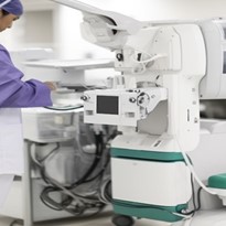
II. Components of a mobile x-ray machine: X-ray tube, detector, and control panel
In this section, we will delve into the fundamental components that constitute a mobile X-ray machine. Understanding these components is essential to comprehend how these machines function and produce diagnostic images with precision.
A. X-ray tube
- Function of the X-Ray Tube: The X-ray tube is at the heart of any X-ray machine, including mobile X-ray machines. Its primary function is to generate X-rays through a process known as X-ray emission. These X-rays are then directed toward the patient's body to create diagnostic images.
- Components of the X-Ray Tube: The X-ray tube consists of several key components, each with a specific role in the X-ray generation process. These components include:
- Cathode: The cathode is the negative electrode within the X-ray tube. It emits a focused electron beam when heated. This electron beam is crucial for initiating X-ray production.
- Anode: The anode, in contrast, is the positively charged electrode. It serves as the target for the electron beam produced by the cathode. When the high-energy electrons from the cathode strike the anode, X-rays are generated.
- Focusing Cup: The focusing cup surrounds the cathode and helps concentrate the emitted electrons into a narrow beam, ensuring a more precise X-ray generation process.
- Target Material: The anode is typically made of a dense metal, such as tungsten, which is chosen for its ability to withstand the high temperatures generated during X-ray production.
- X-Ray Generation: When an operator initiates an X-ray exposure, the cathode heats up, releasing electrons into the tube. These electrons are accelerated towards the anode by the electric potential between the cathode and anode. As the high-speed electrons strike the anode's target material, they release energy in the form of X-rays. This process, known as bremsstrahlung radiation, results in the production of a spectrum of X-ray energies.
B. Detector
- Role of the Detector: The detector is an integral part of the X-ray machine responsible for capturing the X-rays that pass through the patient's body. It plays a pivotal role in converting X-ray energy into a visible image that can be interpreted by healthcare professionals.
- Types of Detectors: There are various types of detectors commonly used in mobile X-ray machines. These include:
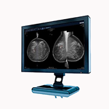
- Film-Based Detectors: Traditional film-based detectors were once widely used but have largely been replaced by digital technology. They involve exposing X-ray film to the transmitted radiation to create a latent image, which is then chemically processed to produce the final X-ray image.
- Digital Detectors: Modern mobile X-ray machines predominantly employ digital detectors. These can be further categorized into two types:
- Computed Radiography (CR): CR systems use a photostimulable phosphor plate to capture X-rays. After exposure, the plate is scanned, and the captured data is processed to create a digital image.
- Direct Radiography (DR): DR systems employ a digital detector panel that directly converts X-rays into electrical signals. This immediate conversion allows for rapid image acquisition and real-time viewing.
C. Control panel
- Functions of the Control Panel: The control panel of a mobile X-ray machine serves as the command center for the operator. It provides a user-friendly interface for adjusting various parameters during the imaging process. Functions of the control panel include:
- Exposure Settings: Operators can select the appropriate X-ray exposure parameters, such as exposure time and tube current, to tailor the imaging process to the patient's needs.
- Image Processing: Some control panels offer image processing features that allow for adjustment of image contrast and brightness post-acquisition.
- Patient Data Entry: Operators can input essential patient information directly into the control panel to associate it with the acquired images.
- Operator's Role: Operators play a critical role in ensuring the accuracy and safety of X-ray imaging by using the control panel to configure settings according to the specific clinical requirements. Proper training is essential to handle this aspect of the mobile X-ray machine effectively.
III. Emitting x-ray beams and capturing images
A. X-ray emission
X-ray beams are generated within the mobile X-ray machine through a process known as X-ray emission. This critical step involves the conversion of electrical energy into X-rays, which are then directed towards the patient for imaging purposes.
Factors Affecting X-ray Beam Intensity:
- Voltage (kVp): The voltage applied to the X-ray tube determines the energy of the emitted X-rays. Higher voltages result in more penetrating X-rays.
- Current (mA): The X-ray tube current regulates the number of X-ray photons produced per unit of time. It affects the overall brightness of the image.
- Exposure Time: The duration for which the X-ray tube is active directly influences the amount of radiation exposure to the patient and the image quality.
B. Patient positioning
Proper patient positioning is of utmost importance in obtaining accurate and diagnostically valuable X-ray images. The positioning of the patient affects the clarity and detail present in the radiographic image.
Importance of Proper Patient Positioning:
- Anatomy Visualization: Correct positioning ensures that the area of interest is in the X-ray beam's path, allowing for optimal visualization of the anatomy being examined.
- Avoiding Artifacts: Incorrect positioning can lead to artifacts, such as blurriness or distortion, which can hinder diagnostic accuracy.
- Minimizing Radiation Exposure: Precise positioning helps focus the X-ray beam on the targeted area, reducing unnecessary exposure to surrounding tissues.
C. Image capture
The process of capturing X-ray images involves the use of a detector to record the X-rays that pass through the patient's body. This recorded information is then used to create a digital or analog image.
Key Aspects of Image Capture:
- Detector Technology: Modern mobile X-ray machines typically use digital detectors, which offer several advantages such as real-time imaging and improved image quality. Older systems may use analog film.
- Radiation Exposure: The X-ray photons that pass through the patient interact with the detector, creating an image. The amount of radiation exposure must be carefully controlled to minimize the potential risks to the patient.
- Image Resolution: Image resolution refers to the level of detail captured in the X-ray image. Higher resolution allows for better visualization of small structures, making it an important consideration in diagnostic imaging.
IV. Real-time imaging and its importance in critical situations
A. Real-time x-ray imaging
Real-time imaging in the context of X-rays refers to the immediate and continuous visualization of internal structures and processes within the human body. Unlike traditional static X-ray images, which provide a single snapshot, real-time X-ray imaging allows healthcare professionals to observe dynamic changes as they occur.
Examples of Critical Situations Where Real-Time Imaging is Crucial:
- Emergency Trauma Care: In trauma situations, such as car accidents or falls, where rapid assessment is critical, real-time X-ray imaging helps in identifying fractures, internal bleeding, and foreign objects in real-time. This enables medical teams to make quick decisions about interventions and surgical procedures.
- Cardiac Catheterization: During cardiac catheterization procedures, real-time X-ray imaging is used to guide catheters through blood vessels to the heart. This continuous imaging assists in diagnosing and treating coronary artery diseases and structural heart problems promptly.
- Interventional Radiology: In interventional radiology procedures, real-time X-ray guidance is essential for tasks like placing stents, removing blood clots, or delivering localized treatments for conditions like tumors. This precise, dynamic imaging ensures that medical interventions are accurate and minimally invasive.
B. Benefits of real-time imaging
Real-time X-ray imaging offers several advantages in the field of healthcare, enhancing both diagnosis and treatment:
- Immediate Feedback: Healthcare professionals can see the effects of their actions in real-time, allowing for quick adjustments during procedures. This immediate feedback improves the precision and safety of medical interventions.
- Reduced Radiation Exposure: While real-time imaging uses X-rays, it typically requires shorter exposure times compared to traditional X-rays. This reduces radiation exposure for both patients and healthcare providers.
- Minimally Invasive Procedures: Real-time imaging enables minimally invasive surgeries and interventions, which often lead to shorter recovery times, reduced pain, and lower risks of complications compared to open surgeries.
- Enhanced Diagnostic Accuracy: Real-time X-ray imaging aids in diagnosing conditions that may be challenging to detect with static images alone. For example, it can reveal the precise location of a needle during a biopsy or the movement of contrast agents in blood vessels.
- Improved Patient Outcomes: The ability to monitor and adjust treatments in real-time can lead to better patient outcomes, particularly in critical situations where time is of the essence.
V. Ensuring safety for patients, operators, and healthcare professionals
A. Radiation safety
Radiation safety is paramount when utilizing X-ray technology. Mobile X-ray machines are designed with multiple safety features to mitigate radiation exposure:
- Lead Shielding: Mobile X-ray machines incorporate lead shielding to contain and minimize radiation exposure to surrounding areas. This ensures that radiation is directed only toward the targeted body part.
- Collimators: Collimators are devices that restrict the X-ray beam to the specific area of interest, reducing unnecessary radiation exposure to adjacent tissues.
- Dosimetry Monitoring: Healthcare facilities employ dosimetry badges to track the radiation exposure of healthcare professionals regularly. This helps ensure that exposure levels remain within safe limits.
B. Operator training
Operators of mobile X-ray machines undergo rigorous training and certification to ensure safe and accurate operation:
- Radiologic Technologists: Radiologic technologists are trained to operate X-ray equipment, including mobile units. They receive education on radiation safety, patient positioning, and the proper use of imaging equipment.
- Certification: In many regions, radiologic technologists must obtain certification or licensure to practice. This certification process includes examinations and assessments of their skills and knowledge.
- Continuing Education: To maintain their certification, operators often must participate in ongoing education and training to stay current with advancements in technology and safety protocols.
C. Patient safety
Ensuring patient safety is a top priority during X-ray procedures:
- Informed Consent: Before any X-ray procedure, patients are provided with detailed information about the procedure's purpose, potential risks, and benefits. Informed consent is obtained to ensure patients understand and agree to the procedure.
- Lead Aprons and Shields: Patients are typically provided with lead aprons or shields to protect areas of their bodies not under examination from unnecessary radiation exposure.
- Pregnancy Screening: It's essential to screen female patients for pregnancy, as radiation exposure during pregnancy can harm the developing fetus. If pregnancy is suspected, alternative imaging methods may be considered.
In conclusion, mobile X-ray machines are invaluable tools in the realm of healthcare, providing medical professionals with a convenient and efficient means of conducting diagnostic imaging. This comprehensive guide has shed light on the intricate workings of these machines, emphasizing their fundamental principles, key components, and applications.