Ultrasound machines have revolutionized medical imaging by providing non-invasive and real-time visualization of internal structures. To maximize the diagnostic potential of ultrasound, a range of accessories and upgrades are available that enhance imaging capabilities and enable specialized applications. In this guide, we will explore the world of ultrasound machine accessories and upgrades, as well as their various applications in medical practice. From transducers and probes to software upgrades, we will delve into the advancements that contribute to improved imaging quality and expanded diagnostic possibilities. Let's dive in and explore the exciting world of ultrasound machine accessories and upgrades.
Ultrasound Machine Accessories and Upgrades
Ultrasound machine accessories and upgrades offer a variety of features and functionalities that enhance imaging performance and broaden diagnostic capabilities. Some key accessories and upgrades include:
1. Transducers and Probes
Transducers and probes are essential accessories that transmit and receive ultrasound waves. They come in various shapes, sizes, and frequencies to accommodate different imaging needs. Higher frequency transducers provide better resolution for superficial imaging, while lower frequency transducers are suitable for deeper structures. Specialized probes, such as endocavity or intracavitary probes, are designed for specific applications, such as transvaginal or transrectal imaging.
2. Software Upgrades
Software upgrades play a crucial role in improving image quality, enhancing user experience, and expanding the functionality of ultrasound machines. Upgrades may include advanced image processing algorithms, automated measurement tools, and new imaging modes. These upgrades allow for better visualization of anatomical structures, improved image clarity, and more efficient workflow.
3. Ergonomic Accessories
Ergonomic accessories focus on enhancing user comfort and ergonomics during ultrasound examinations. These may include adjustable height tables, footrests, and arm supports. By providing better positioning options and reducing strain on the operator, ergonomic accessories contribute to improved image quality and increased operator efficiency.
4. Storage and Connectivity Solutions
Storage and connectivity solutions enable efficient management of ultrasound images and data. These accessories include PACS (Picture Archiving and Communication Systems) integration, DICOM (Digital Imaging and Communications in Medicine) compatibility, and network connectivity options. These features facilitate seamless image transfer, remote viewing, and integration with electronic medical record (EMR) systems, ensuring effective data management and streamlined workflow.
Top Accessories and Add-Ons for Your Ultrasound Machine
Specialized Probes and Transducers
Specialized probes and transducers are among the top accessories that can enhance your ultrasound machine's capabilities. These include:
1. 3D/4D Imaging Probes
3D/4D imaging probes enable the visualization of volumetric data, providing a more comprehensive view of anatomical structures. These probes are particularly useful in obstetrics, gynecology, and fetal imaging, allowing for detailed assessments of fetal development and monitoring.
2. High-Frequency Probes
High-frequency probes offer improved resolution for superficial imaging. They are ideal for applications such as musculoskeletal, vascular, and small parts imaging, where fine details need to be visualized with high precision.
3. Transesophageal Echocardiography (TEE) Probes
TEE probes are specialized probes designed for cardiac imaging. They are inserted into the esophagus to obtain detailed images of the heart and surrounding structures. TEE probes provide enhanced visualization for cardiac assessments and interventions.
4. Endocavity Probes
Endocavity probes are designed for imaging within body cavities, such as the rectum or vagina. These probes are used in urology, gynecology, and prostate imaging, enabling close proximity imaging with higher resolution.
Image Management and Reporting Systems
Efficient image management and reporting systems are essential for streamlining workflow and ensuring accurate documentation. Consider the following accessories:
1. Picture Archiving and Communication System (PACS)
PACS allows for the storage, retrieval, and distribution of medical images. It enables seamless integration with your ultrasound machine, facilitating efficient image management, remote access, and collaborative consultations.
2. Image Analysis and Measurement Software
Image analysis and measurement software provide advanced tools for analyzing ultrasound images. These software solutions offer precise measurements, automated calculations, and quantitative analysis, assisting in accurate diagnosis and monitoring.
3. Reporting Templates and Integration
Reporting templates and integration features streamline the reporting process by providing standardized templates and enabling integration with electronic medical record (EMR) systems. This ensures consistent and comprehensive reporting, improving communication and documentation.
Wireless Connectivity Options
Wireless connectivity options can enhance the flexibility and accessibility of your ultrasound machine. Consider the following accessories:
1. Wireless Transducers
Wireless transducers eliminate the need for cable connections, offering greater freedom of movement during examinations. They allow for more convenient positioning and reduce the risk of cable-related artifacts.
2. Mobile Device Integration
Integration with mobile devices, such as smartphones or tablets, enables remote control and image viewing. This facilitates on-the-go access to images and simplifies communication with colleagues, enhancing collaboration and efficiency.
3. Cloud-Based Storage and Sharing
Cloud-based storage and sharing platforms allow for secure storage, backup and sharing of ultrasound images. This enables easy access to images from multiple devices and locations, promoting seamless collaboration and efficient remote consultations.
Enhanced Visualization and Ergonomics
Enhanced visualization and ergonomics accessories can improve user experience and optimize imaging outcomes. Consider the following additions:
1. Adjustable Monitor Mounts
Adjustable monitor mounts allow for flexible positioning of the ultrasound monitor, ensuring optimal viewing angles and reducing strain on the operator. This improves ergonomics and enhances comfort during long scanning sessions.
2. Anti-Glare Screens
Anti-glare screens reduce reflections and glare on the ultrasound monitor, improving image visibility in bright environments. They enhance image interpretation and reduce eye fatigue during scanning.
3. Contrast Enhancement Technologies
Contrast enhancement technologies, such as adaptive image processing algorithms, improve the visualization of subtle tissue variations and enhance image contrast. These technologies aid in the detection of pathology and improve diagnostic confidence.
Biopsy and Interventional Accessories
For ultrasound-guided biopsies and interventional procedures, specific accessories can enhance precision and procedural success. Consider the following additions:
1. Biopsy Guides and Needle Holders
Biopsy guides and needle holders provide stability and accuracy during ultrasound-guided biopsies. They help maintain the desired needle trajectory, reducing the risk of inadvertent tissue damage and improving sample acquisition.
2. Needle Visualization Technologies
Needle visualization technologies, such as needle tracking or needle enhancement software, enhance the visibility of the biopsy needle during procedures. This aids in accurate needle placement and improves procedural safety and efficiency.
3. Sterile Covers and Probe Protectors
Sterile covers and probe protectors maintain a sterile barrier during invasive procedures, reducing the risk of cross-contamination. These accessories ensure procedural safety and promote infection control measures.
Applications of Ultrasound Accessories and Upgrades
Ultrasound machine accessories and upgrades have broad applications across various medical specialties. Some notable applications include:
1. Obstetrics and Gynecology
Ultrasound plays a crucial role in obstetrics and gynecology for fetal monitoring, assessing pregnancy viability, and diagnosing gynecological conditions. Specialized transducers and probes, such as transvaginal and 3D/4D imaging probes, enable detailed visualization of the fetus and reproductive organs, enhancing diagnostic accuracy.
2. Cardiology
In cardiology, ultrasound accessories and upgrades enable advanced imaging techniques, such as echocardiography and stress echocardiography. Transesophageal probes provide clear visualization of cardiac structures, while contrast-enhanced ultrasound allows for enhanced detection of perfusion abnormalities and assessment of myocardial function.
3. Radiology
In radiology, ultrasound accessories and upgrades expand the diagnostic capabilities of ultrasound imaging. Specialized transducers and software upgrades enable applications such as elastography, which assesses tissue stiffness for the detection of tumors or liver fibrosis. Additionally, contrast-enhanced ultrasound with microbubble agents enhances vascular imaging and aids in the characterization of lesions.
4. Interventional Procedures
Ultrasound-guided interventional procedures benefit from accessories like needle guides and specialized probes. These accessories improve needle visualization and accuracy during biopsies, aspirations, and other interventions. Real-time imaging guidance enhances safety and precision, reducing the risk of complications.
5. Point-of-Care Ultrasound (POCUS)
Point-of-care ultrasound applications benefit from portable ultrasound machines and wireless connectivity options. With the use of handheld devices and mobile applications, healthcare providers can perform focused examinations at the patient's bedside, improving triage decisions, rapid diagnosis, and monitoring of critical conditions.
6. Musculoskeletal Imaging
Ultrasound accessories and upgrades in musculoskeletal imaging offer detailed visualization of muscles, tendons, ligaments, and joints. High-frequency linear probes and specialized software enable dynamic imaging, assessment of tendon integrity, and guided interventions such as injections or aspirations.
7. Urology
In urology, accessories like endocavity probes and software upgrades facilitate the assessment of the urinary system. Transrectal or transvaginal probes aid in the evaluation of the prostate, while advanced imaging techniques like shear wave elastography assist in characterizing renal or bladder lesions.
Types of Ultrasound Probes
Ultrasound probes can be classified into several types based on their design and functionality. The main types include:
1. Linear Array Probes
Linear array probes consist of multiple transducer elements arranged in a line. These probes are ideal for imaging superficial structures, such as blood vessels, tendons, and breasts. They provide high-resolution images and are commonly used in musculoskeletal, vascular, and small parts imaging.
2. Curved Array Probes
Curved array probes have a slightly curved transducer array, allowing for better imaging of deep structures and organs. These probes are commonly used in abdominal, obstetric, and gynecological imaging. The curved design enables improved visualization and easier maneuverability in these areas.
3. Phased Array Probes
Phased array probes utilize multiple elements that can be electronically focused and steered to create images. These probes are particularly useful in cardiac imaging, where they enable the visualization of structures at different angles. Phased array probes are also utilized in abdominal and pediatric imaging.
4. Endocavity Probes
Endocavity probes are designed for imaging within body cavities, such as the rectum or vagina. These probes have a smaller footprint and are equipped with higher frequency transducers for improved resolution. Endocavity probes are commonly used in urology, gynecology, and prostate imaging.
5. Transesophageal Probes
Transesophageal probes are inserted into the esophagus to provide detailed imaging of the heart and surrounding structures. These probes offer high-frequency imaging capabilities and are widely used in cardiac imaging, especially during intraoperative or interventional procedures.
6. Intraoperative Probes
Intraoperative probes are specifically designed for imaging during surgical procedures. These probes are sterilizable and can be used directly on the patient's body during surgery. They provide real-time imaging guidance, assisting surgeons in visualizing critical structures and ensuring precise interventions.
Sizes of Ultrasound Probes
Ultrasound probes are available in various sizes, each suitable for specific applications and imaging depths. Common size classifications include:
1. Small Parts Probes
Small parts probes are used to image superficial structures, such as the thyroid, breast, or testicles. These probes typically have higher frequency transducers, enabling detailed imaging of small and shallow areas.
2. Convex Probes
Convex probes have a wider footprint and are suitable for imaging larger areas, such as the abdomen or obstetric examinations. These probes provide a balance between imaging depth and field of view, making them versatile for various applications.
3. Microconvex Probes
Microconvex probes have a smaller footprint compared to convex probes and are used in applications where space is limited, such as pediatric and neonatal imaging. These probes offer high-resolution imaging in small anatomical areas, providing detailed visualization.
4. Phased Array Probes
Phased array probes come in different sizes and are utilized in various applications, including cardiac and vascular imaging. The size of the phased array probe depends on the specific imaging requirements, such as the depth of the structures being examined.
5. Endocavity Probes
Endocavity probes are available in different sizes to accommodate various anatomical requirements. The size of the endocavity probe typically corresponds to the specific application, such as transvaginal or transrectal imaging in gynecology and urology.
6. Transesophageal Probes
Transesophageal probes come in different sizes to suit patients of different ages and anatomical variations. These probes are designed to be inserted into the esophagus for cardiac imaging, and their size is selected based on the patient's anatomy and the specific imaging needs.
Upgrading Your Ultrasound Machine: Software and Hardware Options
As technology continues to advance, upgrading your ultrasound machine can significantly enhance its performance and expand its capabilities. Upgrades can encompass both software and hardware options, offering improved imaging quality, increased efficiency, and access to advanced features. In this guide, we will explore the various software and hardware options available for upgrading your ultrasound machine. From advanced imaging algorithms to hardware enhancements, these upgrades have the potential to transform your ultrasound system and provide a higher level of diagnostic accuracy. Let's dive in and discover the possibilities of upgrading your ultrasound machine.
Software Upgrades
Software upgrades offer an array of benefits and enhancements to your ultrasound machine. These upgrades typically involve updating the underlying software platform of the system, introducing new features, improving image processing algorithms, and enhancing workflow efficiency. Some common software upgrades include:
1. Advanced Image Processing Algorithms
Upgrading your ultrasound machine's software can introduce advanced image processing algorithms that enhance image quality and diagnostic clarity. These algorithms can improve resolution, reduce image artifacts, and enhance visualization of subtle anatomical structures. By upgrading to the latest image processing software, you can significantly enhance the diagnostic capabilities of your ultrasound system.
2. Workflow Optimization Tools
Software upgrades often include workflow optimization tools that streamline the imaging process, improve efficiency, and reduce user fatigue. These tools can include automated measurement tools, customizable reporting templates, and streamlined image management systems. Upgrading your ultrasound machine's software with these workflow optimization tools can save time, improve productivity, and enhance the overall user experience.
3. New Imaging Modes and Techniques
Software upgrades may introduce new imaging modes and techniques that expand the diagnostic capabilities of your ultrasound system. These can include advanced imaging techniques such as elastography, shear wave imaging, contrast-enhanced ultrasound, and fusion imaging. By upgrading your ultrasound machine's software, you gain access to these cutting-edge imaging modalities, enabling more accurate diagnoses and improved patient care.
Hardware Upgrades
Hardware upgrades involve replacing or enhancing specific hardware components of your ultrasound machine to improve its performance and functionality. These upgrades can offer significant improvements in image quality, system durability, and user experience. Some common hardware upgrades include:
1. Transducer Upgrades
Transducer upgrades involve replacing or adding new transducers to your ultrasound system. Upgrading to higher frequency transducers can improve resolution for superficial imaging, while adding specialized transducers for specific applications can expand the diagnostic capabilities of your ultrasound machine. Transducer upgrades allow for better imaging of different anatomical regions and provide greater flexibility in performing various examinations.
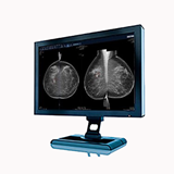
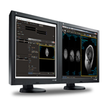
2. Display and Monitor Upgrades
Upgrading the display and monitor of your ultrasound machine can have a significant impact on image visualization and user experience. Upgrading to higher-resolution displays with improved color accuracy and contrast can enhance image clarity and improve diagnostic confidence. Larger and more ergonomic monitors can also improve user comfort and efficiency during examinations.
3. Processing Unit and Memory Upgrades
Upgrading the processing unit and memory of your ultrasound machine can enhance its overall performance and speed. Upgrading to a more powerful processor and increasing the memory capacity can enable faster image acquisition, processing, and storage. This results in smoother workflow, reduced waiting times, and improved efficiency during examinations.
Ultrasound Printers: Types, Features, and Benefits
Ultrasound printers are available in different types, each offering specific features and print capabilities. The main types of ultrasound printers include:
1. Thermal Printers
Thermal printers are the most commonly used printers in ultrasound imaging. They use heat to transfer images onto special thermal paper, eliminating the need for ink or toner cartridges. Thermal printers offer fast printing speeds, high-resolution output, and excellent image clarity. They are cost-effective and produce long-lasting prints that resist fading over time.
2. Color Printers
Color printers provide the ability to print ultrasound images in color, enabling enhanced visualization of anatomical structures and pathology. These printers are particularly useful in applications such as obstetrics, where color-coded images assist in identifying blood flow and differentiating tissues. Color printers offer the advantage of improved diagnostic interpretation and communication.
3. Dye-Sublimation Printers
Dye-sublimation printers utilize a heat transfer process to produce high-quality prints with vibrant colors and continuous tones. These printers are capable of reproducing detailed grayscale images and color images with exceptional accuracy. Dye-sublimation printers are often used in specialized applications where color fidelity and image detail are critical, such as dermatology or plastic surgery.
4. Inkjet Printers
Inkjet printers use liquid ink cartridges to create prints. While less commonly used in ultrasound imaging, inkjet printers offer the advantage of producing high-quality color prints with excellent image detail. They provide the flexibility to print on a variety of paper types and sizes, making them suitable for specific imaging requirements.
Features and Benefits of Ultrasound Printers
Ultrasound printers offer several features and benefits that enhance the imaging workflow and facilitate effective communication. Some notable features and benefits include:
1. High-Quality Image Reproduction
Ultrasound printers are designed to produce high-quality prints with accurate representation of anatomical details, grayscale tones, and color information. This allows healthcare professionals to review images in printed format with confidence, aiding in accurate diagnosis and treatment planning.
2. Fast Printing Speeds
With fast printing speeds, ultrasound printers ensure efficient workflow and minimize waiting time for image retrieval. Rapid printouts enable timely consultations, effective communication with patients, and improved overall efficiency in healthcare settings.
3. User-Friendly Interface
Ultrasound printers are equipped with user-friendly interfaces that allow for easy operation and intuitive navigation. They often feature touchscreens or user-friendly control panels, making it simple for healthcare professionals to select print options, adjust settings, and manage printing tasks.
4. Portable and Compact Designs
Many ultrasound printers are designed to be portable and compact, allowing for flexibility in various clinical settings. These printers can be easily transported between examination rooms or taken to remote locations, enabling immediate access to printed ultrasound images wherever they are needed.
5. Connectivity Options
Modern ultrasound printers offer various connectivity options to facilitate seamless integration with ultrasound systems and picture archiving and communication systems (PACS). They can be connected via wired or wireless connections, enabling efficient image transfer and printing directly from the ultrasound system or an electronic medical record (EMR) system.
6. Print Annotation and Labeling
Ultrasound printers often come equipped with features that allow for easy annotation and labeling of printed images. This includes the ability to add patient information, date and time stamps, measurement data, and other relevant details directly onto the printouts. Such annotations aid in accurate documentation and improve the comprehensiveness of patient records.
7. Image Editing and Optimization Tools
Some ultrasound printers offer built-in image editing and optimization tools that allow for on-demand adjustments to image parameters. This includes brightness, contrast, zoom, and image cropping, ensuring that the printed images accurately represent the desired diagnostic information.
8. Cost-Effectiveness
Ultrasound printers, especially thermal printers, are known for their cost-effectiveness. They require minimal maintenance and do not rely on expensive ink or toner cartridges. Thermal paper used in these printers is readily available and affordable, making it a cost-effective choice for printing ultrasound images.
9. Compliance with Medical Regulations
Ultrasound printers are designed to comply with medical regulations and standards, ensuring the privacy and security of patient data. They often incorporate features such as data encryption and user authentication to safeguard patient information during the printing process.
In conclusion, ultrasound machine accessories and upgrades play a crucial role in enhancing the performance and capabilities of ultrasound systems. With a wide range of accessories and upgrades available, such as specialized probes, software enhancements, ergonomic accessories, storage and connectivity solutions, and more, healthcare professionals can benefit from improved imaging quality, expanded diagnostic possibilities, and enhanced workflow efficiency. These advancements contribute to better visualization of anatomical structures, accurate diagnoses, streamlined image management, and improved patient care across various medical specialties. By exploring the world of ultrasound machine accessories and upgrades, healthcare providers can maximize the potential of ultrasound technology and further revolutionize medical imaging.