
The preanalytic phase (that is, sample collection, handling and transportation) the analytic phase; and the postanalytic phase (that is, transmission and interpretation of the test result, as well as clinical response to the test result in terms of patient management).
Error can occur during all three phases, but study has demonstrated the preanalytic phase to be particularly prone, accounting for up to 70% of all errors in the process of testing patient samples [1].
This is the fourth of a series of articles that together address the issue of preanalytic error - and its avoidance - in blood gas testing.
The three previous articles focused on the non-traditional analytes that are now available on blood gas analyzers, namely: electrolytes [2]; the metabolites, glucose, lactate and creatinine [3]; and bilirubin [4].
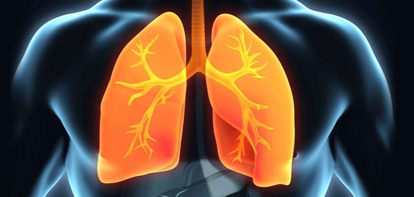
Here the focus is the traditional parameters of blood gas analysis used to assess patient acid-base and oxygenation status: pH, pCO2 and pO2.
For this analyte specific consideration, discussion will be largely concerned with preanalytic factors that might jeopardize preservation of in-vivo values ofpH, pCO2 and pO2.
However, for completeness, the article begins with a brief discussion of the more general errors that can occur during the preanalytic phase of any laboratory (or point-of-care) test, irrespective of the mode of analysis or analyte being measured.
Elements of the preanalytic phase
The preanalytic phase of patient blood testing is a process that begins with test ordering by a clinician, and includes all subsequent steps up to the point the patient sample (either whole-blood, plasma or serum) is analyzed.
Each of these preanalytical steps is associated with the potential for error as outlined in Table 1.
|
PREANALYTICAL STEP |
SOME POTENTIAL ERRORS |
|
1. Test ordered by clinician |
Inappropriate test ordering [5] |
|
2. Identification of patient |
Misidentification - wrong patient tested [6,7] |
|
3. Preparation of patient |
Patient inadequately prepared (e.g. patient not adequately fasted prior to measurement of glucose) |
|
4. Sample collection
|
- Poor blood collection technique causing hemolysis [8] or venostasis - Wrong sample collected or sample collected in wrong tube type - Inadequate mixing of sample with chemicals - anticoagulant, preservative - in collection tube - Inadequately/inaccurately labelled collection tube [6,7] |
|
5. Sample transport |
- transport not sufficiently rapid to ensure sample integrity and/or preservation of in-vivo analyte concentration - temperature during transport may threaten sample integrity and/or preservation of in-vivo analyte concentration - mode of transport may threaten sample integrity and/or preservation of in-vivo analyte concentration |
|
6. Sample reception and preparation for analysis in the laboratory |
|
There is relatively recent acknowledgment that it is sometimes sensible to consider steps 1-5 separately from step 6 because steps 1-5 (dubbed the pre-preanalytic phase) are performed outside the laboratory, largely by non-laboratory staff, whereas step 6 (called the preanalytic phase) is performed within the laboratory, solely by laboratory staff.
Study has shown that errors occur much more frequently in the pre-preanalytic than the preanalytic phase [9].
For this article only the term preanalytic will be used; no distinction will be made between preanalytic and pre-preanalytic.
Preanalytic factors that might affect pH, pCO2 and pO2.
The principle aim of preanalytical quality must be to preserve in-vivo values of pH, pCO2 and pO2 so that measured values most accurately reflect the patient’s acid-base and oxygenation status at the time blood was sampled.
With this aim in mind, the following preanalytical factors will be considered:
- Patient preparation
- Type of blood sample (arterial, venous and capillary)
- Sample collection
- anaerobic technique
- plastic versus glass syringe
- anticoagulation
- Sample handling and transport
- effect of time between sampling and analysis
- temperature of sample during transport
- mode of transport
Patient preparation
The gold standard sample for measurement of pH, pCO2 and pO2 is arterial blood [10]. Sampling of arterial blood is painful and may result in pain/anxiety-induced hyperventilation, which can potentially cause spuriously reduced pCO2.
A calm and reassuring manner during preparation of the patient, along with application of local anesthetic to the proposed sampling site [11] might reduce the risk of this potential artefactual effect on pCO2.
Since administration of oxygen and rate of mechanical ventilation affect measured values, it is important that blood is sampled after a period of stabilization following introduction or dose change of these interventions.
Guidelines [10] suggest this time to steady state is 20-30 minutes, but results of a recent study [12] suggest a delay of only 15 minutes is necessary to assure steady state after administration of oxygen or dose change.
Type of blood sample
Although arterial blood is the preferred ‘gold standard’ sample for measurement of pH, pCO2 and pO2, venous blood collected from either a peripheral vein or central venous line may be an acceptable alternative if only pH and pCO2 (measures of acid-base status) are of interest.
A study has demonstrated that the arterio-venous difference for these two analytes is small and consistent, so that it is possible to predict - with arguably clinically acceptable degree of accuracy - arterial pH and pCO2 from measured venous pH and pCO2 [13].
A recent meta-analysis [14] of 18 previous studies examining the validity of using venous blood concluded that venous blood was suitable for pH measurement, but less suitable for measuring pCO2.
Capillary blood collected by fingertip, heel or earlobe stab is an acceptable alternative sample to arterial blood if only pH and pCO2 are required [15,16].
Whilst there is support for the notion that venous and capillary blood samples are acceptable – though less than perfect – alternatives to arterial blood for measurement of pH and pCO2, there is universal agreement that both are unacceptable for assessing blood oxygenation (pO2).
The arterio-venous difference for pO2 is large and variable [13]. If determination of patient pO2 is required, arterial blood is the only acceptable sample.
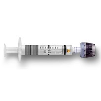
Sample collection – anaerobic technique
In order to preserve in vivo value of pO2, and to a lesser extent pH and pCO2, it is vital that blood is collected and transported without exposure to air (i.e. collected anaerobically).
The requirement that blood is not exposed to air determines that any air bubbles trapped in the blood-filled syringe must be expelled immediately after the sample is collected, and that the syringe is then capped for the duration of period between collection and analysis.
Blood is aspirated directly from syringe to analyzer in order to preserve anaerobic conditions.
These recommendations are based on evidence from a number of studies conducted over the years [17-20] that have demonstrated that equilibration of oxygen between blood and air causes a time-dependent in vitro change in pO2.
Typically, the effect is to increase pO2; however, the issue is more complex. Initial pO2 of the sample determines both the magnitude of the change and the direction of the resulting bias.
pO2 increases if initial pO2 is less than that of ambient air (i.e. ~ 150 mmHg, 20kPa), and decreases if initial pO2 is greater than that of ambient air (the latter case would only, of course, apply if the patient was receiving supplemental oxygen).
The magnitude of the change is not as great for hypoxemic (initial low pO2) samples as it is for normoxemic (initial pO2 in the reference range) samples [20].
Some studies reveal that air contamination can also cause a slight (usually clinically insignificant) increase in pH and decrease in pCO2.
Sample collection - glass versus plastic syringe
The notion that the syringe material (glass or plastic) might affect in vivo values has been studied.
In summary, these studies [21-23] indicate that glass and plastic syringes are equally effective at preserving pH and pCO2, but that glass syringes preserve pO2 values better than plastic syringes.
This difference is likely due to the relatively higher oxygen permeability of plastic [24].
Movement of oxygen from ambient air across the plastic syringe wall to the blood sample can cause an artefactual, time and temperature dependent, increase in pO2.
The potential for error in pO2 measurement due to the permeability of plastic can be eliminated or at least minimized by:
• maintaining the sample at room temperature - rather than at lower temperature (e.g. in iced water), and
• analyzing the sample immediately (or at least within 15-30 minutes of sampling).
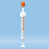
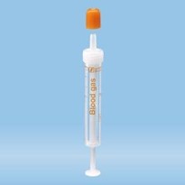
For safety and convenience reasons, commercially available plastic syringes specifically designed for blood-gas testing are the most commonly used, in preference to glass syringes.
A recent comparative study [25] of these commercially available plastic syringes suggests that differences in syringe manufacture is a potential source of preanalytic variation, although this variation was found not to be clinically significant with regard to pH and pO2, but maybe in the case of pCO2 measurement.
Sample collection - anticoagulation
Aspiration of a homogenous whole blood sample into the blood gas analyzer requires that the sample be artificially anticoagulated to prevent in vitro clotting.
Heparin is the only anticoagulant suitable for blood gas testing [26].
Liquid heparin solutions, once used to anticoagulate blood gas samples, are best avoided because their use is associated with risk of over diluting blood samples, which can cause erroneously low pCO2 results [27].
The now recommended alternative is the dried (lyophilized) electrolyte balanced heparin preparations that are present in commercially available plastic blood gas syringes.
In some poorly resourced areas of the world, the relative expense of commercial syringes has determined that locally prepared syringes with liquid heparin continue to be used.
The shortcomings of this practice are highlighted by the results of a recent study [28].
For effective anticoagulation, it is of course essential that the lyophilized heparin is distributed quickly and evenly through the whole blood sample by thorough but gentle mixing.
The recommended mixing technique is a repetitive combination of inverting the syringe several times then rolling it through the palms of the hands. Study suggests that a mixing time of up to 2 minutes is required to restore a cell sedimented sample to homogeneity [29].
Inadequate or delayed mixing can result in inadequate anticoagulation and the formation of fibrin clots in the sample that might block the analyzer and prevent analysis.
Inadequate anticoagulation due to inadequate mixing of the sample remains a common reason for the rejection of blood-gas samples [30].
Although a more vigorous mixing technique would, intuitively at least, promote anticoagulation, it should be avoided because it might cause red blood cell destruction (hemolysis), which can have the effect of reducing pH and pO2 values and increasing pCO2 values [31].
Sample transport – effect of time delay and temperature
As previously mentioned, the in vitro changes in pH, pCO2 and pO2 due to contaminating air and the use of plastic syringes are time dependent.
The longer the period between collection and analysis (i.e. transport time), the greater is the magnitude of these changes.
A quite separate problem of delay in analysis is posed by the fact that blood cells continue to metabolize glucose after collection. This in vitro glycolysis is associated with consumption of oxygen and generation of carbon dioxide.
There is thus a time-dependent artefactual decrease in pO2 and increase in pCO2 due to ongoing in vitro glycolysis [32]. Since glycolysis is an enzyme-mediated process, it is temperature dependent.
The lower the temperature of the sample, the slower in vitro glycolysis proceeds and consequently the slower is the rate of in vitro pO2 decline and pCO2 rise.
This provides the rationale for the once widespread practice of maintaining samples at 0OC. between collection and analysis, by placing the syringe in an iced-water slurry.
Unfortunately, lowering the temperature of the sample (if it is contained within a plastic syringe) has the deleterious effect of increasing oxygen permeability of the syringe, giving rise to artefactual increase in pO2, as described above.
Since the cooling of samples in plastic syringes is contraindicated, because of its effect on pO2, the only way of ameliorating the potential deleterious effect of in vitro glycolysis on pO2 and pCO2 values is to analyze the sample as soon as possible, so that in vitro glycolysis has minimal effect.
Two recent studies [33,34] investigated the effect of delay in analysis and storage temperature of blood for preservation of in vivo pH, pCO2 and pO2 values, and have provided evidence in support of these previously expressed expert recommendations [11,35]:
“If blood is collected into a plastic syringe it should be kept at room temperature and analyzed within 15 minutes if pO2 is required, but within 30 minutes otherwise.
If blood cannot be analyzed within 30 minutes, blood should be collected into, preferably, a glass syringe.
The sample should be placed in iced-water slurry to reduce sample temperature and thereby minimize in vitro glycolysis. The sample should be analyzed within 60 minutes of collection”.
The rate of in vitro glycolysis is a function of the number of metabolically active cells in the blood sample.
This poses a particular problem for preservation of in vivo pO2values among patients with myeloproliferative diseases, such as acute leukemia, that are associated with extremely high white cell (leucocyte) or platelet counts.
For these patients the rate of in vitro glycolysis and consequent rate of in vitro oxygen consumption is so high that it results in spuriously low pO2 unless blood is analyzed immediately after it is sampled (i.e. at the patient’s bedside) [36].
Blood should be sampled into a pre-cooled syringe, kept on ice and analyzed within a few minutes if bedside analysis is not feasible [37].
Sample transport – mode of transport
Pneumatic tube transport (PTS) provides the means for rapid transport of blood-gas samples from the point-of-care to the central laboratory; the alternative is human courier.
A number of studies have examined the potential for PTS to affect in vivo pH, pCO2 and pO2.
A review of these studies [38] concludes that pH and pCO2 are unaffected by PTS, but that pO2 can be affected if the sample is contaminated with even tiny amounts of air.
PTS amplifies the effect of contaminating air on pO2 [18,20] outlined above.
It is thus particularly important to pay scrupulous attention to the removal of air bubbles from arterial blood samples if they are to be transported via PTS.
Summary - the tips
The measurement of pH, pCO2 and pO2 is vulnerable to a number of preanalytic errors.
The following tips are intended to help avoid these errors and thereby ensure that results of analysis accurately reflect the patient’s current acid-base and oxygenation status.
- Positively identify your patient (wrist band and verbal confirmation with patient if conscious)
- Label sample container (syringe) with patient identifying barcode before proceeding to blood collection
- Before collecting blood, allow time (at least 15 minutes) for stabilization following a change in mechanical ventilation rate/oxygen therapy.
- Remember: Arterial blood is the preferred gold-standard sample for measurement of pH, pCO2 and pO2
- Do not consider sampling venous/capillary blood if pO2 is of clinical interest
- Immediately after the blood is sampled, remove any air from the sample by holding the syringe vertically and gently tapping the sides to dislodge trapped air bubbles, then expelling them along with any air in the luer of the syringe to paper tissue (the use of vented caps is recommended for this procedure)
- Cap the syringe immediately after air has been expelled
- Thoroughly, but gently, mix the sample to ensure adequate anticoagulation by repeatedly inverting the capped syringe and rolling it between the palms of your hands
- Analyze the sample immediately, or at least within 15 minutes of collection if pO2 is of interest, otherwise within 30 minutes of collection
- Maintain samples collected in plastic syringes at room temperature (do not ice) for the duration of time between collection and analysis
- If blood cannot be analyzed within 30 minutes, it should ideally be collected into a glass syringe, but whether glass or plastic syringes are used, the collected sample should be stored at 0oC by immersing the syringe in an iced-water slurry. Samples stored in this way must be analyzed within 60 minutes of collection
- Remember: Transport via pneumatic tube is associated with the risk of erroneous pO2 results, if even tiny amounts of air remain in the syringe
Summary
The measurement of the parameters pH, pCO2 and pO2 is vulnerable to a number of preanalytical errors and this article shares practical tips to help avoid these errors, ensuring that the results of analysis accurately reflect the patient’s acid-base and oxygenation status.
The tips include the removal of air bubbles, correct identification of the patient, labeling of the sample container, thorough mixing of the sample and analysis within 15 minutes of sample collection.
-160x160-state_article-rel-cat.png)