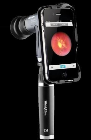
Welch Allyn, a leading medical diagnostics device manufacturer that specialises in improving patient outcomes, today announced it has utilised advanced ophthalmology technology already in the hands of physicians to create the Welch Allyn iExaminer™—a product that consists of a hardware adapter and associated software that allows healthcare providers to capture, store, send and retrieve images from the Welch Allyn PanOptic™ Ophthalmoscope using the iPhone™ 4 or 4S.
The PanOptic features patented optical technology that creates a viewing area of the fundus and retinal nerve in an undilated pupil that is 5 times larger than that of a traditional ophthalmoscope and increases magnification by 26 percent to more easily see retinal details.
The iExaminer rapidly captures and transmits the retinal images created by the PanOptic for easy, cost-effective eyeground image documentation.
"The iExaminer allows healthcare providers to easily capture and share the images of a fundus in a moment's notice, helping to improve the quality of care provided—especially for remote users who may not have easy access to specialists," said Rick Farchione, senior manager, physical assessment at Welch Allyn.
"It will increase workflow efficiency by allowing providers to capture and share images from any clinical environment. It is a low-cost way to digitally capture eye imaging and will also make it easier for providers to share images with their patients, helping to improve patient knowledge and compliance."
The iExaminer adapter aligns the optical access of the PanOptic to the visual axis of the iPhone 4 or 4S camera to capture high resolution pictures of a patient's fundus and retinal nerve.
The iExaminer software application, available from the Apple App Store, then allows physicians to save images to a patient file, as well as e-mail and print the images.
"This is the first affordable device to give almost anyone, anywhere the ability to capture a picture of the back of the eye," said Dr. Wyche Coleman, inventor of the iExaminer. "I was able to take this very lightweight, portable, inexpensive iExaminer to the summit of Mount Kilimanjaro in sub-Saharan Africa and take a picture of a patient's fundus.
From the top of the mountain, I then transmitted it to a doctor at Johns Hopkins University in the United States where he was able to analyse the image."


