Radiation safety and compliance are paramount considerations in the realm of X-ray machines, where accurate diagnostics must harmonize with meticulous protection measures. Within the purview of medical imaging, dental diagnostics, and various industrial applications, X-ray machines play an indispensable role in revealing internal structures and aiding diagnoses. This introduction delves into the foundational importance of adhering to regulatory standards, ensuring the safety of both operators and patients, and upholding impeccable radiation safety practices.
I. Ensuring Compliance with Regulatory Standards
A. Overview of Key Regulatory Bodies
- The Food and Drug Administration (FDA) and the International Atomic Energy Agency (IAEA) stand as pivotal players in the formulation and enforcement of safety standards for X-ray machines.
- The FDA, within the United States, provides guidelines that encompass device performance, labeling, and quality control, with the primary goal of guaranteeing patient safety.
- The IAEA, on an international scale, establishes frameworks that promote the responsible and safe use of radiation-emitting equipment, fostering a global culture of radiation safety.
B. Significance of Adhering to Standards
- Adherence to regulatory standards is non-negotiable when it comes to X-ray machine operation.
- These standards are meticulously crafted to minimize radiation exposure risks for patients, healthcare workers, and the general public.
- By strictly following these guidelines, healthcare facilities and professionals mitigate potential health hazards and enhance the overall quality of patient care.
C. Discussion of Potential Consequences
- Non-compliance with regulatory standards can result in a range of adverse outcomes.
- Legal ramifications, including fines and potential facility closures, may be imposed due to violations.
- More critically, failure to adhere to safety standards can lead to increased radiation exposure, compromising the health of both patients and healthcare personnel.
- Additionally, reputational damage and loss of public trust can ensue, impacting the credibility of healthcare institutions.
II. Radiation Shielding Measures for Operator and Patient Safety
A. Explanation of the Principles of Radiation Shielding and Its Importance
- Radiation shielding refers to the use of materials and techniques to minimize the exposure of individuals to ionizing radiation.
- The primary goal of shielding is to block or absorb harmful radiation, thus reducing the risk of health hazards associated with prolonged or high-dose exposure.
- Shielding operates on the basis of attenuation, where the shielding material absorbs or scatters radiation, preventing it from reaching operators and patients.
B. Different Types of Shielding Materials and Techniques Used in X-ray Machine Design:
- Lead Shielding: Lead is a commonly used material due to its high density and effective radiation absorption properties. It is often integrated into the walls, doors, and protective barriers of X-ray rooms.
- Concrete Shielding: Concrete, particularly high-density concrete, is utilized in constructing walls and partitions to create a barrier against radiation.
- Gypsum Board: Gypsum board with lead lining is employed to enhance radiation protection in walls and ceilings.
- Lead Aprons and Protective Clothing: Operators wear lead aprons and other shielding garments to minimize radiation exposure to sensitive body areas during procedures.
C. Emphasis on How Proper Shielding Enhances Safety for Both Operators and Patients:
- Operator Safety: Adequate shielding safeguards healthcare workers, such as radiologic technologists, from unnecessary radiation exposure during daily tasks. This reduces their risk of developing radiation-related health issues over time.
- Patient Safety: Shielding mechanisms within X-ray machines ensure that the patient is exposed only to the necessary amount of radiation required for accurate diagnostics. This helps prevent overexposure and associated risks.
- Collateral Exposure Minimization: Shielding not only protects direct users but also minimizes radiation leakage, thereby safeguarding individuals in nearby areas.
III. Monitoring Radiation Exposure for Healthcare Workers
In a healthcare environment where X-ray machines are utilized for diagnostics and treatments, the continuous monitoring of radiation exposure among healthcare workers becomes paramount. This practice aligns with the stringent emphasis on radiation safety and compliance, aiming to safeguard both medical professionals and patients. To ensure adherence to radiation exposure limits and maintain a safe working environment, the implementation of effective monitoring measures is essential.
-
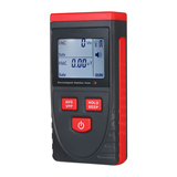
The Role of Dosimeters: Tracking Cumulative Radiation Doses
Dosimeters, specialized devices worn by healthcare workers, play a pivotal role in tracking the cumulative radiation doses they are exposed to during their duties. These devices are designed with precision to measure ionizing radiation, offering a comprehensive overview of the actual radiation levels experienced over time. Dosimeters can be worn on the body or placed in the working environment, allowing for accurate data collection in various medical scenarios.
-
Importance of Data Collection and Trend Identification
Collecting and analyzing dosimeter data is not only a compliance requirement but also a proactive strategy to identify trends and potential hazards. By closely monitoring the radiation exposure patterns of healthcare workers, medical facilities can identify high-exposure areas, time frames, or procedures. Such insights enable the formulation of informed protocols to reduce exposure and enhance safety measures.
-
Ensuring Safe Radiation Levels
The data collected from dosimeters serve as a foundation for ensuring safe radiation levels within the healthcare setting. By comparing the recorded radiation doses against established regulatory limits and best practice guidelines, medical institutions can assess compliance and make necessary adjustments. In instances where exposure levels approach or exceed recommended limits, corrective actions can be swiftly taken to minimize risk.
IV. Implementing Protocols to Minimize Unnecessary Radiation Exposure
-
Explanation of the ALARA (As Low As Reasonably Achievable) Principle
The ALARA principle stands as a cornerstone in the realm of radiation safety and X-ray procedures. Its fundamental premise is simple yet crucial: radiation exposure should be maintained "As Low As Reasonably Achievable," while still achieving the necessary diagnostic information. This principle underscores the commitment to minimizing radiation doses without compromising the quality of medical imaging.
-
Description of Protocols and Best Practices for Minimizing Radiation Exposure during X-ray Procedures
To implement the ALARA principle effectively, a series of protocols and best practices have been developed to guide healthcare professionals during X-ray procedures. These protocols are grounded in both scientific research and practical experience, ensuring that radiation exposure is reduced to the lowest levels possible without compromising patient care. Key components of these protocols include:
Patient Assessment and Justification: Before initiating any X-ray procedure, a thorough evaluation of the necessity of the examination is crucial. Healthcare professionals must ensure that the benefits of the procedure outweigh the potential risks associated with radiation exposure. This step involves clinical judgment and shared decision-making with the patient.
Optimized Technique Selection: The choice of X-ray technique plays a pivotal role in radiation exposure. Professionals should select techniques that balance diagnostic accuracy with minimal radiation output. Modern X-ray machines often offer a variety of exposure settings that can be tailored to the specific needs of the patient and the area being imaged.
Collimation and Shielding: Proper collimation focuses the X-ray beam only on the area of interest, minimizing unnecessary exposure to surrounding tissues. Additionally, shielding with lead aprons and other protective devices is vital to protect non-imaged areas of the body and personnel present during the procedure.
Use of Digital Imaging: Digital radiography and fluoroscopy systems offer significant advantages over traditional film-based methods. They require lower radiation doses to produce high-quality images and provide real-time visualization, allowing for shorter exposure times.
Avoiding Repeat Exposures: Repeat exposures should be minimized whenever possible. Ensuring patient cooperation and proper positioning during the initial exposure can reduce the need for additional imaging attempts.
Regular Equipment Calibration and Maintenance: Periodic calibration and maintenance of X-ray equipment are essential to ensure that the machine functions optimally. Properly calibrated equipment produces consistent and accurate images with lower radiation doses.
-
Benefits of Optimized Exposure for Accurate Diagnostics and Reduced Health Risks
Implementing protocols to minimize radiation exposure offers a myriad of benefits, primarily centered around accurate diagnostics and decreased health risks for both patients and healthcare workers.
Accurate Diagnostics: Optimized exposure protocols result in high-quality images that facilitate precise diagnosis. Clearer images allow healthcare professionals to make accurate and timely clinical decisions, leading to better patient outcomes.
Reduced Health Risks: Minimizing radiation exposure is directly linked to lowering the potential health risks associated with ionizing radiation. By adhering to ALARA principles and employing best practices, the likelihood of radiation-induced complications, such as tissue damage and the development of secondary cancers, is significantly diminished.
Enhanced Patient Safety: Patients can be reassured that medical professionals prioritize their safety. Transparent communication about the necessity of X-ray procedures, along with visible safety measures such as shielding and proper collimation, fosters a sense of trust between patients and healthcare providers.
Long-term Radiation Safety for Healthcare Workers: Minimizing radiation exposure benefits not only patients but also healthcare workers who perform X-ray procedures regularly. Adhering to stringent safety protocols ensures that medical personnel are exposed to minimal radiation doses over their careers, reducing the risk of occupational hazards.
V. Proper Disposal of Hazardous Materials and Waste
Proper disposal of hazardous materials and waste generated by X-ray machines is an essential component of radiation safety and compliance. It not only safeguards the environment but also prevents potential health risks associated with the mishandling of radioactive substances. This section delves into the importance of appropriate disposal methods, outlines protocols for safe waste management, and explores considerations related to environmental impact and health hazards.
A. Importance of Proper Disposal of Materials Contaminated by Radiation
Materials exposed to radiation can become contaminated with radioactive particles or substances. Improper disposal of these materials poses risks not only to the environment but also to individuals who might come into contact with them. Contaminated waste can spread radiation, leading to long-term health consequences and environmental degradation. Proper disposal ensures that the radiation exposure is limited and contained, mitigating potential harm.
B. Explanation of Protocols for Safe Disposal of Hazardous Waste Generated by X-ray Machines
- Segregation and Identification: Waste generated during X-ray procedures needs to be segregated based on its level of radioactivity. This segregation enables proper categorization and ensures that waste is treated in accordance with its potential risks. Radioactive waste is typically classified into low-level, intermediate-level, and high-level waste categories.
- Packaging and Labeling: Once segregated, waste should be appropriately packaged and labeled to indicate its radioactive nature and associated hazards. Proper labeling assists waste handlers in identifying and managing the waste safely. Packaging also prevents leakage and contamination during transportation and storage.
- Storage: Before disposal, radioactive waste is often stored temporarily to allow for decay of short-lived radioactive isotopes. Storage facilities must adhere to strict safety standards and regulations to prevent any release of radiation into the environment. Regular monitoring and inspection ensure the integrity of these storage facilities.
- Treatment and Disposal: Different types of radioactive waste require distinct treatment and disposal methods. Low-level waste, which includes materials with minimal radioactivity, can often be safely disposed of in designated landfills. Intermediate and high-level waste, which pose higher risks, may require specialized treatment, such as encapsulation or deep geological disposal.
C. Considerations for Environmental Impact and Potential Health Hazards
Environmental Impact: Improper disposal of radioactive waste can have lasting negative impacts on the environment. Contaminated materials can seep into soil and water sources, affecting ecosystems and potentially entering the food chain. Adhering to proper disposal protocols helps minimize the environmental footprint of radiation-related activities.
- Health Hazards: Radioactive waste poses serious health hazards if not managed properly. Individuals who come into contact with inadequately handled waste can experience radiation exposure, leading to increased cancer risks, genetic mutations, and other health issues. Following disposal protocols mitigates these risks and ensures the safety of both workers and the general public.
- Regulatory Compliance: Regulatory bodies such as the Nuclear Regulatory Commission (NRC) enforce stringent guidelines for the disposal of radioactive waste. Compliance with these regulations is paramount to prevent legal repercussions and to maintain the trust of the public in the healthcare industry's commitment to safety.
Conclusion
In conclusion, the landscape of X-ray machines in medical diagnostics presents a tapestry woven with the threads of radiation safety and compliance. The symbiotic relationship between technological advancement and ethical responsibility underscores the paramount importance of adhering to regulatory standards and implementing stringent safety measures. By upholding the principles of radiation shielding, monitoring exposure, and minimizing unnecessary risks, the healthcare community can ensure the well-being of both patients and operators. This discourse sheds light on the multifaceted nature of this vital topic, urging stakeholders to remain vigilant in their pursuit of excellence while embracing the ever-evolving landscape of medical imaging. As we delve deeper into the subsequent sections, a more comprehensive panorama of the intricacies and imperatives of radiation safety and compliance will unfold.