According to Dr Jinman Kim, team leader in the Institute of Biomedical Engineering and Technology (BMET), medical imaging is now a fundamental aspect of healthcare delivery but the challenge facing clinicians is how best to extract or identify relevant information from these massive data sets.
Working with clinicians based at the Royal Prince Alfred hospital, the team has already developed a 3D 'virtual human body', programmable to an individual's medical history and viewable on mobile devices.
The group was recently awarded an Australian Research Council (ARC) grant to expand its work in this area and develop methods of extracting and harnessing knowledge from large collections of biomedical imaging data.
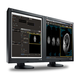
"These data come from a variety of imaging scanners used to acquire images of the human body, such as dual-modal positron emission tomography and computed tomography (PET-CTs), ultrasound, and magnetic resonance imaging (MRIs)," explains Dr Kim.
"Existing imaging datasets store vital historical diagnostic information that can provide clinicians with an evidence-based resource to assist in their patient diagnosis and research.
"But manual identification, labelling, and analysis of tumours from these images can be time consuming," he says.
The new ARC funded project will build on the work of PhD graduate Ashnil Kumar whose research findings were published in this month's journal of medical image analysis and investigated graph-based techniques for searching medical images based on patterns described by shape, colour, texture, and the spatial arrangement of objects.
Ashnil states: "We hypothesised that the spatial mapping of different tumours could provide important insights into the heterogeneous manifestations of the disease and its patterns of spreading.
"Our algorithm took this theory into account particularly in the case of patients with non-small cell lung cancer (NSCLC)."
The ARC funded project will provide opportunities for content-based image search and retrieval, case-based reasoning, training, and comparison of data from clinical trials.

- Suppliers
- New to MedicalSearch? Book a Demo
- Advertise with us
- Login
- Email Marketing
- Buyers
- Get Quotes
- Articles & Ideas
- Login
- Subscribe to newsletter
- My Details
- Get Quotes
- Accident & Emergency Care
- Aged Care & Disability
- Anaesthesia & Respiratory Care
- Beauty & Wellness
- Cardiology & Cardiac Surgery
- Commercial Cleaning & Laundry Supplies
- Dental Care & Oral Surgery
- Diagnostic Instruments & Medical Imaging
- Disinfection & Sterilisation
- ENT & Audiology
- Gynaecology & Obstetrics
- Homecare & Consumer Medical
- Hospital Equipment & Supplies
- Intensive Care Unit
- Laboratory & Pathology
- Medical Apparel
- Medical Devices & Products
- Medical Fridges & Freezers
- Medical Storage & Filing
- Medical Waste Management
- Optometry & Ophthalmology
- Orthopaedics & Podiatry
- Paediatrics & Neonatology
- Patient Monitoring & Management
- Physiotherapy & Rehabilitation
- PPE & Infection Control
- Single Use Medical Consumables
- Surgical Tools & Supplies
- Treatment Beds, Tables & Couches
- Veterinary Equipment
- Wheelchairs & Mobility Aids
Trusted by 520,000 Australian medical buyers
Buyers
- Discover products & solutions
- Login
- Subscribe To Newsletter
- Browse All Products
- Read Articles
Suppliers
Advertise
- Promote your products & solutions
- New to MedicalSearch? Book a Demo
- Login / Forgot Password
- Advertise Your Products
- Success Stories
- Email Marketing
- Suppliers
- Advertise with us
- Login
- Email Marketing
- Buyers
- Get Quotes
- Articles & Ideas
- Login
- Subscribe to newsletter
- My Details
Get Quotes
- Accident & Emergency Care
- Aged Care & Disability
- Anaesthesia & Respiratory Care
- Beauty & Wellness
- Cardiology & Cardiac Surgery
- Commercial Cleaning & Laundry Supplies
- Dental Care & Oral Surgery
- Diagnostic Instruments & Medical Imaging
- Disinfection & Sterilisation
- ENT & Audiology
- Gynaecology & Obstetrics
- Homecare & Consumer Medical
- Hospital Equipment & Supplies
- Intensive Care Unit
- Laboratory & Pathology
- Medical Apparel
- Medical Devices & Products
- Medical Fridges & Freezers
- Medical Storage & Filing
- Medical Waste Management
- Optometry & Ophthalmology
- Orthopaedics & Podiatry
- Paediatrics & Neonatology
- Patient Monitoring & Management
- Physiotherapy & Rehabilitation
- PPE & Infection Control
- Single Use Medical Consumables
- Surgical Tools & Supplies
- Treatment Beds, Tables & Couches
- Veterinary Equipment
- Wheelchairs & Mobility Aids
Trusted by 520,000 Australian medical buyers