Creating histopathology sections is a crucial aspect of the field of pathology, enabling pathologists to examine tissue samples in detail to diagnose diseases, study cell structures, and investigate the effects of various treatments. Histopathology sections provide valuable insights into the cellular and tissue-level changes that occur in the body. In this article, we will explore the process and equipment used in the creation of histopathology sections, shedding light on the techniques and tools that are essential for this vital medical practice.
What is Histopathology?
Histopathology is the branch of pathology that focuses on the microscopic examination of tissue samples. It plays a pivotal role in the diagnosis and treatment of various medical conditions, including cancer, infectious diseases, autoimmune disorders, and more. The histopathology process involves the preparation of tissue samples and their subsequent examination under a microscope to study cellular morphology, tissue architecture, and any pathological changes.
Importance of Histopathology
Histopathology is a cornerstone of modern medicine. It helps clinicians and pathologists make accurate diagnoses, predict disease outcomes, and determine the most suitable treatment options. Without histopathology, the understanding and management of diseases would be severely limited.
The following are some key aspects of histopathology:
Disease Diagnosis: Histopathology allows for the identification of diseases at an early stage, facilitating prompt treatment.
Treatment Planning: By examining tissue samples, pathologists can recommend the most effective treatment options, such as surgery, chemotherapy, or radiation therapy.
Cancer Staging: For cancer patients, histopathology helps determine the extent of the disease, aiding in the staging of cancer and guiding treatment decisions.
Research and Education: Histopathology is vital for medical research and education, enabling scientists and students to explore tissue samples and develop a better understanding of diseases and their mechanisms.
To achieve accurate and reliable results in histopathology, a well-defined process and specialized equipment are essential.
The Histopathology Process
The creation of histopathology sections involves several critical steps, each of which contributes to the quality and accuracy of the final analysis. Here, we outline the key stages of the histopathology process:
Tissue Collection: The first step in histopathology is obtaining tissue samples, which can be collected through various means, such as biopsies, surgical resections, or autopsies. The collection process must ensure the preservation of tissue integrity.
Tissue Fixation: To prevent tissue degradation and maintain its structure, the collected tissue samples are placed in a fixative solution, commonly formalin. This step stabilizes the tissue and prepares it for further processing.
Tissue Processing: After fixation, tissue samples go through a series of dehydration, clearing, and infiltration steps to remove water and replace it with paraffin wax, which makes the tissue suitable for sectioning.
Embedding: The dehydrated and cleared tissue is embedded in paraffin wax, forming a solid block that can be easily sliced into thin sections. This block is known as a paraffin block.
Sectioning: The paraffin-embedded tissue block is cut into extremely thin sections (typically 4-5 micrometers thick) using a microtome. These sections are mounted onto glass slides for further processing.
Staining: Staining is a crucial step in histopathology that helps highlight specific tissue structures and cellular components. Hematoxylin and eosin (H&E) staining is the most commonly used staining method, providing contrast between cell nuclei and the surrounding cytoplasm.
Mounting and Coverslipping: Once the sections are stained, they are mounted on glass slides and covered with a thin, transparent glass coverslip to protect the tissue and maintain its integrity.
Microscopic Examination: The prepared histopathology slides are examined under a light microscope by a pathologist or a trained technician. This examination involves studying the tissue structure, cell morphology, and any abnormalities or disease-related changes.
Diagnosis and Reporting: Based on the microscopic examination, the pathologist makes a diagnosis and prepares a report that includes their findings and recommendations. This report is crucial for patient management and further treatment decisions.
Now that we've outlined the essential steps in the histopathology process, let's delve into the equipment used at each stage.
Equipment for Creating Histopathology Sections
The process of creating histopathology sections involves a range of specialized equipment to handle tissue samples effectively, ensure their preservation, and enable accurate microscopic examination. Here, we'll discuss the key equipment used at each stage of the histopathology process:
1. Tissue Collection:
Biopsy Instruments: These instruments are used to obtain tissue samples through various methods, including core needle biopsies, fine needle aspirations, and excisions. Biopsy needles, forceps, and brushes are commonly used tools.
Surgical Instruments: In surgical resections and autopsies, a variety of instruments such as scalpels, scissors, and retractors are used to collect tissue samples safely and efficiently.
2. Tissue Fixation:
Formalin: Formalin is a fixative solution that contains formaldehyde, used to preserve the structural integrity of tissue samples. It crosslinks proteins and nucleic acids, preventing decomposition.
Tissue Cassettes: These small containers hold tissue samples during fixation. They are usually made of plastic and have small compartments to hold individual samples.
3. Tissue Processing:
Tissue Processor: A tissue processor automates the steps of dehydration, clearing, and infiltration by sequentially immersing the tissue samples in various solutions. It ensures consistent and precise processing.
4. Embedding:
Paraffin Wax: Paraffin wax is used to embed dehydrated tissue samples, creating solid blocks. The blocks are then sliced into thin sections for mounting on slides.
Embedding Station: An embedding station includes a heated paraffin bath and molds for creating paraffin blocks. It allows the embedding process to be performed systematically.
5. Sectioning:
Microtome: A microtome is a precision instrument used to cut thin sections of tissue from paraffin blocks. It is equipped with a knife or blade that can be adjusted to control section thickness.
Microtome Blades: High-quality microtome blades are essential for achieving thin and consistent sections. They must be sharp and maintained properly.
6. Staining:
Staining Stations: Specialized staining stations are used to apply stains to tissue sections. These stations may include multiple staining dishes and a slide rack for efficient staining.
Stains: Various stains are used in histopathology, with hematoxylin and eosin (H&E) being the most common. Hematoxylin stains nuclei blue, while eosin stains cytoplasm and other structures pink.
7. Mounting and Coverslipping:
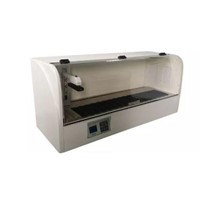
Slide Warmer: A slide warmer is used to help adhere tissue sections to glass slides during the mounting process. It also helps evaporate excess moisture.
Mounting Medium: A mounting medium, typically a clear resin, is applied to the section to secure it to the glass slide. It also prevents air bubbles and provides transparency.
Coverslippers: Automatic coverslippers are used to apply glass coverslips to mounted sections, protecting them and providing a clear view for microscopy.
8. Microscopic Examination:
Microscope: A high-quality light microscope is essential for the examination of histopathology slides. It should have multiple objective lenses, a bright light source, and an adjustable stage for precise focusing.
Camera and Imaging System: Many modern microscopes are equipped with digital cameras and imaging systems that allow for the capture and storage of high-resolution images of the tissue sections.
9. Diagnosis and Reporting:
Computer and Software: Pathologists use computers and specialized software for documenting their findings, preparing reports, and storing digital images for reference.
Pathology Report Templates: Pathologists often use predefined templates to create standardized pathology reports that include patient information, specimen details, findings, and recommendations.
Challenges and Advances in Histopathology
Histopathology is a dynamic field that continually evolves with technological advancements. In recent years, there have been several notable developments and challenges in histopathology:
Digital Pathology: The transition to digital pathology is changing the way pathologists work. Whole-slide imaging allows for the digitization of entire histopathology slides, making it easier to share, store, and analyze images.
Automation: Automated slide scanners and staining systems have improved the efficiency and accuracy of histopathology processes. They reduce human error and increase throughput.
Molecular Pathology: Molecular techniques, such as immunohistochemistry and fluorescence in situ hybridization (FISH), have become essential tools in histopathology, enabling the analysis of specific biomarkers and genetic changes.
Telepathology: Telepathology allows pathologists to remotely access and collaborate on cases, which is particularly valuable in underserved or remote areas.
Quality Control: Quality control measures, including laboratory accreditation and proficiency testing, are critical to ensuring accurate and reliable histopathology results.
Conclusion
Histopathology plays a vital role in the diagnosis, treatment, and understanding of various diseases. The creation of histopathology sections involves a well-defined process, from tissue collection to microscopic examination, and relies on a range of specialized equipment to ensure accurate and reliable results.
Advancements in digital pathology, automation, and molecular techniques are transforming the field, enhancing its efficiency and diagnostic accuracy. The continued progress in histopathology techniques and equipment will undoubtedly contribute to improved patient care and further insights into the complex world of cellular pathology. As technology continues to advance, histopathology will remain at the forefront of medical practice, serving as an invaluable tool in the fight against disease.


-205x205.jpg)
-205x205.jpg)
