
X-ray CT System Full Body CENARIA View
The 64ch/128slice CT system provides images with excellent visibility - even at low doses.
Intelli IPV
Our iterative reconstruction method, Intelli IPV*1, provides images which maintain their natural texture even at high noise reduction rates and their excellent visibility even at low doses, and does not require a dedicated operation room or additional hardware.
Furthermore, by applying a low dose, the anode heat capacity is equivalent to 45 MHU (converted value).
*1 IPV stands for Iterative Progressive reconstruction with Visual modeling.
*2 Scanning the lung field
While adjusting the texture at a uniform ratio from high frequency to low frequency, the physical properties that affect visibility are made as close as possible to FBP.
Visual Model
The Visual Model is a technology that controls image noise and image quality by iterative processing based on statistical model, object model, and physics model.
Object Model
Models morphological information changes to control the size, shape and position of a structure.
Physics Model
The Physics Model is a technology that uses FBP as a model and reduces noise while adjusting the texture from high frequencies to low frequencies at a uniform ratio, achieving a texture close to that of FBP.
Statistical Model
Noise reduction is performed by statistically evaluating X-ray-derived noise and circuit system noise.
Improvement of Low-contrast Detectability
Intelli IPV significantly reduces noise and improves low-contrast detectability while maintaining image texture quality especially in the head region where low-contrast detectability is important.
IntelliEC Plus (combination of auto exposure control and iterative reconstruction)
The tube current is continuously modulated in 3D direction to obtain an image that achieves the target SD. When modulating the tube current, it is possible to set scanning conditions that optimize not only conventional iterative processing but also Intelli IPV characteristics.
SynergyDrive
Examination time is greatly reduced by optimizing the workflow during scanning.
The operator's workflow has been improved by shortening time-consuming procedures.
- Patient positioning
- Patient registration
- Scanogram
- Scanning
- Exit
- Postprocessing
Simultaneous presetting and image processing for multiple patients
The operator is required to perform various tasks in scanning and image processing.
"Study Tab" allows you to switch worksheets for up to 3 patients simultaneously, like flipping pages.
"Navigation Tab" allows you to freely move from scanning to image processing within the same worksheet. Tab switching contributes to a stress-free operator environment.
- Study Tab:"Study Tab"allows easy switching through each patient worksheet.
- Navigation Tab:"Navigation Tab" allows easy switching between imaging and image processing of the same patient.
- Scanning
- Exit
- Image processing
Lateral slide table function for easy positioning
Since the patient table moves laterally up to 200mm (+/-100mm), off-set regions such as the heart and shoulders can be easily positioned at the center of the FOV, improving examination efficiency.
AutoPose (Automatic scan range setting)
Improved reproducibility and reduced set up time.
SCENARIA View can automatically set the scanning range using the scanogram image. The operator can adjust the range manually, if necessary. AutoPose supports several reference lines setting, such as OM Line (Orbitomeatal base line), SM Line (Superior orbitomeatal line) and RB Line (Reid’s base line), for head scanning. Additionally, since the limits can be set in advance, the scan range can be customized according to the preference of the facility.
To contact Fujifilm about X-ray CT System Full Body CENARIA View use Get a quote.
Intelli IPV
Our iterative reconstruction method, Intelli IPV*1, provides images which maintain their natural texture even at high noise reduction rates and their excellent visibility even at low doses, and does not require a dedicated operation room or additional hardware.
Furthermore, by applying a low dose, the anode heat capacity is equivalent to 45 MHU (converted value).
*1 IPV stands for Iterative Progressive reconstruction with Visual modeling.
*2 Scanning the lung field
While adjusting the texture at a uniform ratio from high frequency to low frequency, the physical properties that affect visibility are made as close as possible to FBP.
Visual Model
The Visual Model is a technology that controls image noise and image quality by iterative processing based on statistical model, object model, and physics model.
Object Model
Models morphological information changes to control the size, shape and position of a structure.
Physics Model
The Physics Model is a technology that uses FBP as a model and reduces noise while adjusting the texture from high frequencies to low frequencies at a uniform ratio, achieving a texture close to that of FBP.
Statistical Model
Noise reduction is performed by statistically evaluating X-ray-derived noise and circuit system noise.
Improvement of Low-contrast Detectability
Intelli IPV significantly reduces noise and improves low-contrast detectability while maintaining image texture quality especially in the head region where low-contrast detectability is important.
IntelliEC Plus (combination of auto exposure control and iterative reconstruction)
The tube current is continuously modulated in 3D direction to obtain an image that achieves the target SD. When modulating the tube current, it is possible to set scanning conditions that optimize not only conventional iterative processing but also Intelli IPV characteristics.
SynergyDrive
Examination time is greatly reduced by optimizing the workflow during scanning.
The operator's workflow has been improved by shortening time-consuming procedures.
- Patient positioning
- Patient registration
- Scanogram
- Scanning
- Exit
- Postprocessing
Simultaneous presetting and image processing for multiple patients
The operator is required to perform various tasks in scanning and image processing.
"Study Tab" allows you to switch worksheets for up to 3 patients simultaneously, like flipping pages.
"Navigation Tab" allows you to freely move from scanning to image processing within the same worksheet. Tab switching contributes to a stress-free operator environment.
- Study Tab:"Study Tab"allows easy switching through each patient worksheet.
- Navigation Tab:"Navigation Tab" allows easy switching between imaging and image processing of the same patient.
- Scanning
- Exit
- Image processing
Lateral slide table function for easy positioning
Since the patient table moves laterally up to 200mm (+/-100mm), off-set regions such as the heart and shoulders can be easily positioned at the center of the FOV, improving examination efficiency.
AutoPose (Automatic scan range setting)
Improved reproducibility and reduced set up time.
SCENARIA View can automatically set the scanning range using the scanogram image. The operator can adjust the range manually, if necessary. AutoPose supports several reference lines setting, such as OM Line (Orbitomeatal base line), SM Line (Superior orbitomeatal line) and RB Line (Reid’s base line), for head scanning. Additionally, since the limits can be set in advance, the scan range can be customized according to the preference of the facility.
To contact Fujifilm about X-ray CT System Full Body CENARIA View use Get a quote.
Calculating...
- Your delivery location?
- NSW
- VIC
- QLD
- SA
- WA
- NT
- ACT
- TAS
- New Zealand
- Papua New Guinea
- Afghanistan
- Albania
- Algeria
- Andorra
- Angola
- Antigua and Barbuda
- Argentina
- Armenia
- Austria
- Azerbaijan
- Bahamas
- Bahrain
- Bangladesh
- Barbados
- Belarus
- Belgium
- Belize
- Benin
- Bhutan
- Bolivia
- Bosnia and Herzegovina
- Botswana
- Brazil
- Brunei
- Bulgaria
- Burkina Faso
- Burma
- Burundi
- Cabo Verde
- Cambodia
- Cameroon
- Canada
- Central African Republic
- Chad
- Chile
- China
- Colombia
- Comoros
- Congo (Brazzaville)
- Congo (Kinshasa)
- Costa Rica
- Côte d'Ivoire
- Croatia
- Cuba
- Cyprus
- Czechia
- Denmark
- Djibouti
- Dominica
- Dominican Republic
- Ecuador
- Egypt
- El Salvador
- Equatorial Guinea
- Eritrea
- Estonia
- Ethiopia
- Fiji
- Finland
- France
- Gabon
- Gambia
- Georgia
- Germany
- Ghana
- Greece
- Grenada
- Guatemala
- Guinea
- Guinea-Bissau
- Guyana
- Haiti
- Holy See
- Honduras
- Hungary
- Iceland
- India
- Indonesia
- Iran
- Iraq
- Ireland
- Israel
- Italy
- Jamaica
- Japan
- Jordan
- Kazakhstan
- Kenya
- Kiribati
- Korea, North
- Korea, South
- Kosovo
- Kuwait
- Kyrgyzstan
- Laos
- Latvia
- Lebanon
- Lesotho
- Liberia
- Libya
- Liechtenstein
- Lithuania
- Luxembourg
- Macedonia
- Madagascar
- Malawi
- Malaysia
- Maldives
- Mali
- Malta
- Marshall Islands
- Mauritania
- Mauritius
- Mexico
- Federated States of Micronesia
- Moldova
- Monaco
- Mongolia
- Montenegro
- Morocco
- Mozambique
- Namibia
- Nauru
- Nepal
- Netherlands
- New Zealand
- Nicaragua
- Niger
- Nigeria
- Norway
- Oman
- Pakistan
- Palau
- Panama
- Papua New Guinea
- Paraguay
- Peru
- Philippines
- Poland
- Portugal
- Qatar
- Romania
- Russia
- Rwanda
- Saint Kitts and Nevis
- Saint Lucia
- Saint Vincent and the Grenadines
- Samoa
- San Marino
- Sao Tome and Principe
- Saudi Arabia
- Senegal
- Serbia
- Seychelles
- Sierra Leone
- Singapore
- Slovakia
- Slovenia
- Solomon Islands
- Somalia
- South Africa
- South Sudan
- Spain
- Sri Lanka
- Sudan
- Suriname
- Swaziland
- Sweden
- Switzerland
- Syria
- Taiwan
- Tajikistan
- Tanzania
- Thailand
- Timor-Leste
- Togo
- Tonga
- Trinidad and Tobago
- Tunisia
- Turkey
- Turkmenistan
- Tuvalu
- Uganda
- Ukraine
- United Arab Emirates
- United Kingdom
- United States
- Uruguay
- Uzbekistan
- Vanuatu
- Venezuela
- Vietnam
- Yemen
- Zambia
- Zimbabwe
- NSW
- VIC
- QLD
- SA
- WA
- NT
- ACT
- TAS
- New Zealand
- Papua New Guinea
- Afghanistan
- Albania
- Algeria
- Andorra
- Angola
- Antigua and Barbuda
- Argentina
- Armenia
- Austria
- Azerbaijan
- Bahamas
- Bahrain
- Bangladesh
- Barbados
- Belarus
- Belgium
- Belize
- Benin
- Bhutan
- Bolivia
- Bosnia and Herzegovina
- Botswana
- Brazil
- Brunei
- Bulgaria
- Burkina Faso
- Burma
- Burundi
- Cabo Verde
- Cambodia
- Cameroon
- Canada
- Central African Republic
- Chad
- Chile
- China
- Colombia
- Comoros
- Congo (Brazzaville)
- Congo (Kinshasa)
- Costa Rica
- Côte d'Ivoire
- Croatia
- Cuba
- Cyprus
- Czechia
- Denmark
- Djibouti
- Dominica
- Dominican Republic
- Ecuador
- Egypt
- El Salvador
- Equatorial Guinea
- Eritrea
- Estonia
- Ethiopia
- Fiji
- Finland
- France
- Gabon
- Gambia
- Georgia
- Germany
- Ghana
- Greece
- Grenada
- Guatemala
- Guinea
- Guinea-Bissau
- Guyana
- Haiti
- Holy See
- Honduras
- Hungary
- Iceland
- India
- Indonesia
- Iran
- Iraq
- Ireland
- Israel
- Italy
- Jamaica
- Japan
- Jordan
- Kazakhstan
- Kenya
- Kiribati
- Korea, North
- Korea, South
- Kosovo
- Kuwait
- Kyrgyzstan
- Laos
- Latvia
- Lebanon
- Lesotho
- Liberia
- Libya
- Liechtenstein
- Lithuania
- Luxembourg
- Macedonia
- Madagascar
- Malawi
- Malaysia
- Maldives
- Mali
- Malta
- Marshall Islands
- Mauritania
- Mauritius
- Mexico
- Federated States of Micronesia
- Moldova
- Monaco
- Mongolia
- Montenegro
- Morocco
- Mozambique
- Namibia
- Nauru
- Nepal
- Netherlands
- New Zealand
- Nicaragua
- Niger
- Nigeria
- Norway
- Oman
- Pakistan
- Palau
- Panama
- Papua New Guinea
- Paraguay
- Peru
- Philippines
- Poland
- Portugal
- Qatar
- Romania
- Russia
- Rwanda
- Saint Kitts and Nevis
- Saint Lucia
- Saint Vincent and the Grenadines
- Samoa
- San Marino
- Sao Tome and Principe
- Saudi Arabia
- Senegal
- Serbia
- Seychelles
- Sierra Leone
- Singapore
- Slovakia
- Slovenia
- Solomon Islands
- Somalia
- South Africa
- South Sudan
- Spain
- Sri Lanka
- Sudan
- Suriname
- Swaziland
- Sweden
- Switzerland
- Syria
- Taiwan
- Tajikistan
- Tanzania
- Thailand
- Timor-Leste
- Togo
- Tonga
- Trinidad and Tobago
- Tunisia
- Turkey
- Turkmenistan
- Tuvalu
- Uganda
- Ukraine
- United Arab Emirates
- United Kingdom
- United States
- Uruguay
- Uzbekistan
- Vanuatu
- Venezuela
- Vietnam
- Yemen
- Zambia
- Zimbabwe
Calculating...
Fujifilm X-ray System | FDR Visionary Suite
X-ray System | FDR Smart X
Fujifilm Mammography System | Amulet Sophinity
Fujifilm Ultrasound System | ARIETTA 50
Fujifilm Ultrasound System | ARIETTA 750 DeepInsight
Fujifilm Ultrasound System | ARIETTA 50LE
Fujifilm Ultrasound System | ARIETTA 750SE
Ultrasound System | ARIETTA 650 DeepInsight
Fujifilm Ultrasound System | ARIETTA 65
Fujifilm MRI Scanner | Aperto Lucent
Fujifilm CT Scanner |Supria 32
CT Scanner | SCENARIA View
X-ray CT System Full Body CENARIA View
Mobile X-ray Machine | FDR Go PLUS
Fujifilm DR Detector | FDR D-EVO GL
Fujifilm DR Detector | FDR D-EVO III C25i
Fujifilm CT Scanner |Supria 32
Fujifilm Portable X-ray | FDR Xair
Mobile X-ray Machine | FDR nano
Vet Point-Of-Care Chemistry Analyser | DRI-CHEM NX600V
CT Scanner | SCENARIA View
CT System | Rembra RT CT simulator
CT System | CT 3500
CT System | Spectral CT 7500
CT System | Areta RT CT simulator
CT Scanners | Aquilion ONE / GENESIS Edition
CT Scanners | Aquilion Prime SP
CT Scanners | Aquilion Lightning 160
CT Scanners | Aquilion LB
CT Scanner | SOMATOM Force
CT Scanner | SOMATOM Drive
CT Scanner | SOMATOM X.cite
CT Scanner | 5GXL CBCT
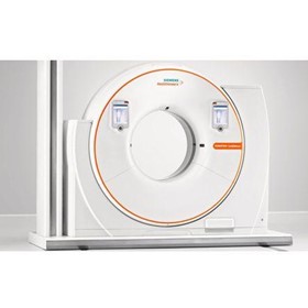
CT Scanner | SOMATOM CT Sliding Gantry
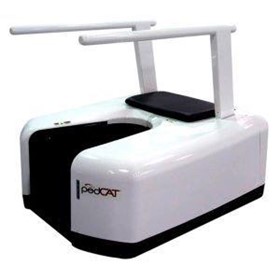
CT Scanner | PedCAT
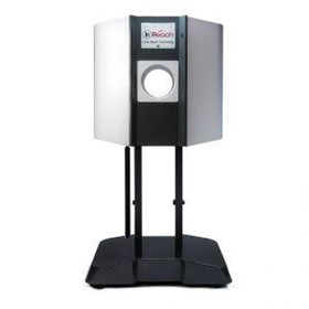
CT Scanner | InReach
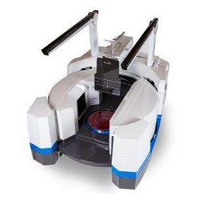
CT Scanner | LineUP
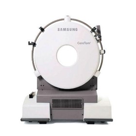
CT Scanners | CereTom
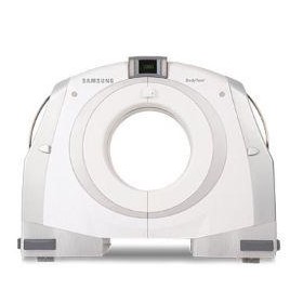
CT Scanners | BodyTom
Thanks for your help!
Sorry! Unable to submit your request at this time. Please try again later.
Report this product